
Chicken Infectious Anemia (CIAV): Symptoms, Pathology & Breeder Immunity
Comprehensive veterinary guide covering Chicken Anemia Virus (Gyrovirus) molecular pathogenesis, thymic atrophy, bone marrow aplasia, and breeder immunization protocols.
Scientific Overview
Chicken infectious anemia is an immunosuppressive viral disease of young chickens caused by Chicken Anemia Virus (CAV / CIAV), a small single-stranded DNA virus belonging to the genus Gyrovirus within the family Anelloviridae. CIAV selectively targets and destroys hemopoietic precursor stem cells in the bone marrow and thymocytes in the cortex of the thymus. The clinical disease causes profound aplastic anemia, subcutaneous hemorrhaging known as blue wing disease, severe immunosuppression, and high secondary bacterial mortality. Understanding chicken anemia virus transmission and breeder seroconversion is essential to protect broiler and layer progeny.
Etiology & Gyrovirus Molecular Architecture
Viral Profile: Chicken Anemia Virus is an unenveloped icosahedral virus containing a circular, negative-sense single-stranded DNA genome of approximately 2.3 kb. CIAV belongs to the family Anelloviridae (genus Gyrovirus) and exhibits extreme resistance to chemical disinfectants, surviving heat at 70°C (158°F) for 1 hour and pH values from 3.0 to 9.0.
The viral genome contains three overlapping open reading frames encoding three essential viral proteins:
- VP1 (51.6 kDa): The major structural capsid protein that bears the neutralizing epitopes. Variations in the hypervariable domain of VP1 modulate viral replication rates and cell tropism.
- VP2 (24.0 kDa): A scaffolding and dual-specificity protein phosphatase required for proper VP1 assembly and structural stability.
- VP3 / Apoptin (13.6 kDa): A potent pro-apoptotic oncolytic protein that induces selective, p53-independent apoptosis in dividing avian lymphoid and hemopoietic stem cells while sparing mature, resting erythrocytes.
Chicken Infectious Anemia Symptoms & Clinical Manifestations
Clinical chicken infectious anemia symptoms manifest predominantly in chicks between 10 and 28 days of age that contracted the virus vertically from non-immune breeder dams lacking maternal neutralizing antibodies. When chicks contract the virus horizontally after 2 weeks of age, clinical anemia is generally absent, but subtle immunosuppression persists:
| Organ System / Feature | Clinical Signs & Pathological Findings | Diagnostic Threshold / Severity |
|---|---|---|
| Hematology (Blood) | Watery, thin, pale blood that clots slowly; severe pancytopenia (reduction in erythrocytes, thrombocytes, and granulocytes) | Packed Cell Volume (PCV / Hematocrit) <20% (normal baseline: 30% to 35%) |
| Integument (Skin) | Severe pallor of comb, wattles, eyelid margins, and shanks; extensive subcutaneous ecchymoses | Pathognomonic "Blue Wing Disease" when complicated by secondary bacterial gangrene |
| General Flock Demeanor | Severe depression, ruffled feathers, huddling under brooders, anorexia, and stunted growth | Flock body weight depression of 15% to 25% below commercial standards |
| Flock Mortality Curve | Mortality begins around Day 12 to 14, peaks between Day 18 and 22, and recedes by Day 28 | Flock mortality typically reaches 5% to 15% (or >30% if complicated by secondary colibacillosis) |
The syndrome known as Blue Wing Disease occurs when profound thrombocytopenia and capillary fragility lead to extensive hemorrhaging beneath the skin of the wings. Opportunistic skin bacteria, particularly Staphylococcus aureus and Clostridium perfringens, invade these oozing hematomas, creating necrotizing, dark purple or blue-black moist gangrenous dermatitis.
Necropsy Pathology: Bone Marrow Aplasia & Thymic Atrophy
Post-mortem dissection of birds affected by Chicken Anemia Virus reveals signature macro-pathological lesions:
- Bone Marrow Aplasia: Splitting the femur lengthwise reveals the most pathognomonic lesion of CIAV: the normal deep red, active hemopoietic marrow is completely replaced by pale yellow, fatty, or translucent pink aplastic tissue.
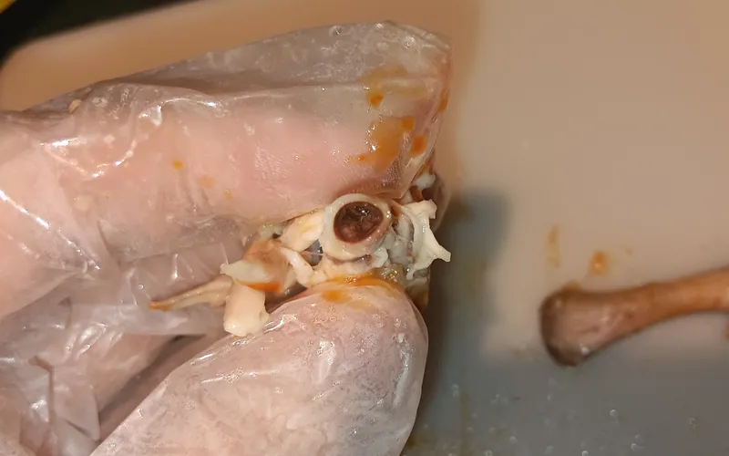
- Thymic Lobular Atrophy: The paired chains of thymic lobes running alongside the jugular veins in the neck shrink dramatically. In advanced cases, individual lobes regress to tiny, dark brown fibrous remnants due to Apoptin-mediated cortical thymocyte lysis.
- Bursal and Splenic Regression: The Bursa of Fabricius and spleen decrease noticeably in size and weight, although bursal atrophy is less intense than in acute Gumboro disease.
- Proventricular Hemorrhages: Petechial and suffusive hemorrhages are frequently visible across the proventricular mucosa and beneath the serosa of skeletal muscles.
Transmission Dynamics: Vertical Shedding vs Horizontal Spread
Epidemiological control of chicken anemia virus requires understanding its twin transmission pathways:
1. Vertical Transmission (The Primary Clinical Threat): Vertical transmission occurs when SPF or unvaccinated breeder pullets enter egg production without prior natural exposure to CIAV. If the naive breeder flock becomes infected during lay (between 25 and 45 weeks of age), dams undergo transient viremia lasting 3 to 6 weeks. During this window, the virus transmits through the fertile egg albumen and yolk to embryos. Hatched chicks lack maternal antibodies, allowing unchecked viral replication in hemopoietic stem cells and triggering clinical aplastic anemia at 12 to 18 days of life.
2. Horizontal Transmission: In older broilers and pullets, transmission occurs via the fecal-oral route through contaminated litter, drinking water, and feather dander. Because birds older than 3 weeks possess age-related immunocompetence, horizontal infection rarely causes clinical anemia; however, it impairs T-cell cytokine synthesis, reducing vaccine efficacy against Newcastle disease and Infectious Bronchitis.
Breeder Immunization & Prevention Protocols
Because no antiviral treatment exists for infected chicks, flock protection relies completely on guaranteeing uniform maternal immunity in breeder parents:
| Control Measure | Recommended Timing | Administration Route | Clinical Objective |
|---|---|---|---|
| Live Attenuated CAV Vaccine | 13 to 15 weeks of age (at least 6 weeks before onset of lay) | Drinking water or wing-web puncture | Induce lifelong seroconversion before fertile egg collection begins |
| Pre-Lay ELISA Serology Audit | 18 to 20 weeks of age | Blood serum sampling (minimum 30 birds per flock) | Confirm 100% flock seropositivity with uniform protective titer titers (CV <30%) |
| Secondary Antimicrobial Support | During clinical outbreaks in broilers (Day 14 to 21) | Drinking water (Amoxicillin or Lincomycin) | Control secondary bacterial gangrenous dermatitis and colibacillosis |
Vaccinating breeder hens later than 6 weeks before lay is strictly avoided to prevent vaccine-strain virus shedding into hatching eggs. High maternal antibodies transferred into the yolk sac protect chicks throughout their first 3 weeks of life, successfully neutralizing early environmental virus challenges.
 biotech Clinical Pathology
Related Calculators
Scientific Literature
- Schat, K. A., & van Santen, V. L. (2020). Chicken Anemia Virus and Other Gyroviruses. In Diseases of Poultry (14th ed., pp. 296-324). Wiley-Blackwell.
- Balamurugan, V., & Kataria, J. M. (2006). Economically important non-oncogenic immunosuppressive viral diseases of chicken: Current status. Veterinary Research Communications, 30(5), 541-566.
- Andrabi, M., et al. (2024). Important non-oncogenic immunosuppressive viral diseases of chickens. Veterinary Research Bulletin, 48(1), 1-18.
- World Organisation for Animal Health (WOAH). (2023). Manual of Diagnostic Tests and Vaccines for Terrestrial Animals.