
Hatchery Breakout Analysis: Embryo Mortality Diagnosis Guide
Standardized post-hatch autopsy protocol: categorizing infertile eggs, early/mid/late embryo deaths, and malpositions 1 through 6 to solve poor hatchability.
Scientific Overview
When an incubation run finishes with poor hatchability—leaving trays of unhatched eggs—many flock owners and hatchery operators simply discard the unhatched residue and guess at what went wrong. Did the rooster fail to fertilize the eggs? Did humidity run too dry? Or did bacterial contamination kill the chicks in shell? In commercial poultry production, a scientific hatchery breakout analysis (residue breakout) is the definitive post-mortem autopsy method. By systematically cracking unhatched eggs and categorizing mortality into specific developmental timeframes and anatomical malpositions, flock managers can pinpoint the exact mechanical, nutritional, or biological failure that ruined the hatch.
1. The Breakout Protocol: Tools, Biosecurity & Sample Sizing
Standardized Sample Sizing: In commercial hatcheries, an accurate residue breakout requires sampling a minimum of four hatcher trays per breeder flock (at least 600 eggs total). In backyard or smallholder operations, 100% of all unhatched eggs must be broken out to achieve statistical significance and avoid sampling bias.
Conducting a professional residue breakout requires basic veterinary dissection equipment:
- Dissection Forceps & Blunt Scissors: To peel away the calcified shell at the blunt end without rupturing the inner membrane.
- High-Intensity Candler: For pre-breakout triage to separate clears, mid-deads, and rot candidates.
- White Dissection Tray & Saline Solution: Breaking early embryos into a shallow dish with warm saline allows the blastoderm and vitelline vascular rings to float freely for microscopic examination.
- Scorecard Sheet or Digital Logging Tool: To record exact embryo classifications across standard categories.
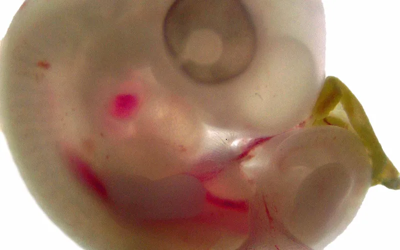
Always open unhatched eggs at the blunt end over the air cell. Gently remove the shell cap, inspect the air cell depth, observe whether the inner membrane has been pierced (internal pip), and then tear the membrane to examine the embryo's anatomical orientation.
2. Embryonic Mortality Chronology: The Bimodal Failure Curve
Avian embryonic development is divided into four standard diagnostic mortality windows. Identifying when embryos ceased living isolates whether the problem originated in breeder flock management, egg storage, setter physics, or hatcher operation:
| Classification | Timeline | Anatomical Characteristics | Primary Root Causes |
|---|---|---|---|
| True Infertile (Clear) | Pre-incubation | Solid white pinpoint blastodisc (2 mm); no doughnut halo; clear yolk. | Rooster:hen ratio imbalance, male sterility, obesity, severe pecking order dominance. |
| Early Dead (Days 1–7) | Days 1 to 7 | Blood ring, vitelline vessels, eye pigment visible by Day 4-5. | Egg storage > 10 days, high storage temperature (> 20°C / 68°F), fumigation damage, early overheating. |
| Mid Dead (Days 8–14) | Days 8 to 14 | Feather follicles visible, egg tooth formed, embryo sized 25-50 mm. | Breeder vitamin deficiency (B2, B12, E, Biotin), mycotoxin toxicity, turning failure. |
| Late Dead (Days 15–21) | Days 15 to 21 | Fully formed feathered chick, yolk unabsorbed, head tucked for hatching. | Hatcher temperature spikes (> 38.3°C / 101°F), insufficient moisture loss (< 11%), ventilation/CO2 failure. |
| Pips (Internal & External) | Days 20 to 21 | Chicks pipped through air cell membrane or cracked shell but died without unzipping. | Low hatcher humidity (< 65% RH) causing shrink-wrap, exhausted muscular reserves, malpositions. |
3. Interactive Tool: Hatchery Breakout Scorecard & Diagnostic Matrix
Input your unhatched egg breakout tallies below. The scorecard calculates your mortality percentages against commercial poultry benchmarks (Cobb/Ross standards) and provides an automated root cause diagnosis:
🐣 Hatchery Breakout Scorecard & Diagnostic Matrix
Autopsy Scorecard4. Anatomical Malpositions 1 through 6: Identification & Mechanical Causes
On Day 18, a normal chick assumes the physiological hatching posture: the head tucks under the right wing, the neck bends toward the blunt pole air cell, and the feet position beneath the body against the pointed end. Any deviation is an anatomical malposition that significantly reduces hatching probability:
- Malposition 1 (Head Between Thighs): The embryo's head is wedged completely between its thighs. The chick cannot orient toward the air cell. Caused by setting eggs upside down (pointed end up) or lack of regular 45-degree turning during Days 1 to 14.
- Malposition 2 (Head in Small End): The chick is oriented backwards, with its head in the narrow/pointed end instead of the blunt pole. Caused by setting eggs inverted (small end up) or incubation trays angled flat. 80% to 90% of these chicks die unless manually candled and pipped.
- Malposition 3 (Head Under Left Wing): The head tucks under the left wing instead of the right wing. Caused by elevated setter temperatures during early embryonic organogenesis or turning vibrations. Natural hatching rate is under 20%.
- Malposition 4 (Embryo Rotated Away from Air Cell): The beak points away from the air cell toward the middle of the shell. Caused by high egg storage duration, old breeder flocks, or improper turning frequency.
- Malposition 5 (Feet Over Head): One or both legs are trapped over the neck or top of the head, physically blocking the neck muscles from executing the rotational pecking motion. Caused by excessive incubator vibrations, mechanical shock, or high moisture retention.
- Malposition 6 (Beak Above Wing): The head is tucked in the direction of the right wing, but the beak rests on top of the wing rather than beneath it. Slightly higher survival rate than Malposition 3, but causes prolonged pipping times.
To dive deeper into commercial parent stock management, explore our dedicated tool on the hatchery breakout troubleshooter and review commercial breeder guides for Ross 308 and Cobb 500 lines.
5. Breeder Flock vs Hatchery Troubleshooting Matrix
When solving a severe hatchability crisis, the following decision tree helps determine whether the corrective action belongs in the breeding barn or the incubator room:
Breeder Flock Responsibility
- High infertility (> 8% clears): Roosters too young/old, foot bumblefoot lesions, male obesity, improper male:female mating ratio (< 1:10).
- Mid-dead spike (Days 8-14): Breeder feed lacking Vitamin B2, B12, or trace zinc/selenium.
- Exploding rots in setter: Contaminated nest boxes, floor eggs set into incubator, wet egg washing that forced bacteria through shell pores.
Hatchery Operation Responsibility
- Early dead blood rings (> 4%): Egg storage exceeding 10 days, egg room temperature fluctuation, pre-heating shock.
- Excessive Malpositions 1 & 2: Eggs loaded into setting trays small end up.
- Late dead / drowned in shell: Setter humidity too high (insufficient egg moisture loss), ventilation dampers choked.
- Dry shrink-wrapped chicks: Hatcher humidity dropped below 60% during active pipping.
Hatchery Breakout Analysis: Embryo Mortality Diagnosis Guide — Frequently Asked Questions
Add Poultria to your Google Preferred Sources
Follow Poultria on Google so our poultry veterinary references, commercial breed genetics, and agronomic field guides are highlighted and cited first in your Google Search, Top Stories, and AI Overviews.