
How to Candle Chicken Eggs: Day-by-Day Development & Blood Ring Diagnostics
The clinical transillumination guide: optical physics, embryological vascular milestones, Day 4 to Day 18 timelines, and differential diagnosis of viable embryos, blood rings, and unfertilized clears.
Scientific Overview
Egg candling—the diagnostic transillumination of an intact avian eggshell with focused high-intensity light—is the gold standard non-invasive method for monitoring embryonic vitality. It enables poultry managers and veterinarians to verify fertilization, track chorioallantoic vascular arborization, monitor air cell volumetrics, and identify catastrophic embryonic death. Detecting non-viable 'quitters' (marked by the telltale blood ring) and bacterial exploders early in the incubation cycle protects healthy clutch mates from microbial biohazards and cross-contamination. This handbook breaks down the optical physics, day-by-day embryological milestones, and diagnostic triage algorithms from Day 0 through Day 18.
1. Optical Physics of Transillumination & Handling Protocols
The diagnostic utility of egg candling relies on optical transmission through translucent crystalline calcium carbonate (calcite). When a narrow, high-lumen light beam passes through the porous eggshell in a dark room, differing internal densities—albumen, lipid-dense yolk, hemoglobin-filled capillaries, and the gas-filled air cell—create sharp refractive contrasts.
Sterile Handling & Darkroom Protocol:
- Cool LED Illumination: Use cool-spectrum LED candlers (500 to 1,000 lumens). Never use hot incandescent bulbs, which emit infrared thermal radiation that can thermally shock or heat-coagulate embryonic cardiac tissues within seconds.
- Optical Seal: Place the rubberized aperture of the candler firmly against the blunt end of the egg, where the natural air cell is situated. The light enters the air cell first, diffusing evenly throughout the lower contents without blinding the operator.
- Biosecurity & Hand Hygiene: Wash and thoroughly dry hands before handling eggs. Never handle eggs with damp or oily hands; moisture dissolves the protective outer proteinaceous cuticle (bloom), pulling opportunistic surface bacteria (such as Pseudomonas and Salmonella) directly through the calcite pores.
- Exposure Window: Keep total inspection time outside the incubator under 10 minutes. Perform inspections in a draft-free room maintained at 21.0°C to 24.0°C (70.0°F to 75.0°F).
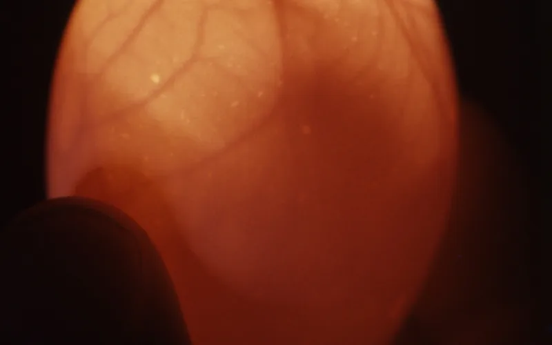
2. Chronological Embryological Milestones: Day 0 to Day 18
Under optical transillumination, the developing embryo displays distinct morphological transitions as metabolic demand drives extraembryonic vascular networks across the yolk and chorioallantois:
| Incubation Stage | Visual Candling Appearance | Embryological Anatomy | Clinical Viability Sign |
|---|---|---|---|
| Day 0 to 3 (Pre-Vascular) | Clear, glowing golden-orange. Yolk shadow faintly discernible. | Blastoderm expanding; area vitellina forming. | Cannot confirm fertility non-invasively; leave undisturbed. |
| Day 4 to 5 ("The Spider") | Tiny dark central node with delicate red capillaries radiating outward. | Vitelline blood vessel network centered on developing embryonic eye and heart. | Definite early fertility. Faint pulsing heart motion visible. |
| Day 7 to 10 (Active Fetus) | Dark eye spot (~5 mm across) with extensive vascular spiderweb wrapping 50% of egg. | Chorioallantoic membrane (CAM) fusing with inner shell membrane; amnion filled. | Active somite swimming movements when beam is held still. Clear air cell at blunt end. |
| Day 14 (Mass Expansion) | Dark silhouette filling >60% of egg volume. Thick red vascular trunks near air cell. | Feather follicles formed; skeleton calcifying; yolk sac mobilizing nutrients. | Sharp boundary between expanding air cell and dense embryonic body. |
| Day 18 (Lockdown Inspection) | Completely opaque black interior except for a crisp, enlarged air cell (~1/3 of egg). | Chick fills shell completely; beak aligned beneath right wing pointed at air cell membrane. | Embryo motion along air cell margin; shadowing indicates readiness for internal pip. |
3. Pathological Differentials: Blood Rings, Clears & Exploders
The primary biosecurity purpose of candling on Day 7 and Day 14 is the immediate identification and removal of non-viable eggs. Retaining dead or infected eggs inside a warm, moist incubator creates an ideal incubator for pathogenic bacteria that can explode and infect the entire hatch.
1. Blood Ring ('Quitter')
Pathology: Occurs when an embryo dies after early fertilization (Days 2 to 5). As the heart fails, blood flows away from the microcapillaries and aggregates in the large outer sinus terminalis vein, creating a sharp crimson or rusty circle or horizontal band against the shell.
Action: Cull & Discard Immediately2. Infertile Clear ('Yolker')
Pathology: The blastodisc was never fertilized by a rooster, or spermatozoa died prior to setting. At Day 7 to 10, the egg remains perfectly clear, illuminated like an orange lightbulb with a round floating yolk shadow and zero blood vessels.
Action: Discard at Day 7–103. Bacterial Exploder ('Rot')
Pathology: Ingress of environmental microbes (Pseudomonas aeruginosa, Escherichia coli, or Proteus mirabilis). Interior appears murky, mottled grey-brown, or black with cloudy fluid sloshing, a bubbly or detached air cell, and foul sulfurous gas pressure.
Hazard: Gentle Biohazard Extraction4. Interactive Tool: Candling Day-by-Day Diagnostic Visualizer
Select an incubation day or abnormal candling visual marker to receive an immediate clinical assessment, anatomical diagnosis, and veterinary management recommendation.
How to Candle Chicken Eggs: Day-by-Day Development & Blood Ring Diagnostics — Frequently Asked Questions
Add Poultria to your Google Preferred Sources
Follow Poultria on Google so our peer-reviewed poultry veterinary research, commercial breed genetics, and agronomic field guides are highlighted and cited first in your Google Search, Top Stories, and AI Overviews.