
Water Belly in Chickens: Ascites Causes, Symptoms & Care
Clinical poultry guide to ascites syndrome: pulmonary hypertension syndrome (PHS), right ventricular congestive heart failure, abdominocentesis, and supportive management.
Scientific Overview
Water belly (clinically termed avian ascites syndrome or Pulmonary Hypertension Syndrome - PHS) is the progressive pathological accumulation of non-inflammatory serous transudate fluid within the coelomic (peritoneal) cavity of domestic poultry. While predominantly recognized in fast-growing broiler meat birds due to extreme metabolic oxygen demands outstripping anatomical pulmonary capillary capacity, ascites frequently afflicts aging backyard laying hens secondary to hepatic lipidosis, salpingitis, chronic respiratory disease, or coelomic adenocarcinoma. Understanding the hemodynamic cascade—from pulmonary vascular resistance to right-sided congestive heart failure and hepatic transudation—is vital for instituting palliative supportive care and making informed animal welfare determinations.
1. Pathophysiology: The Pulmonary Hypertension Cascade
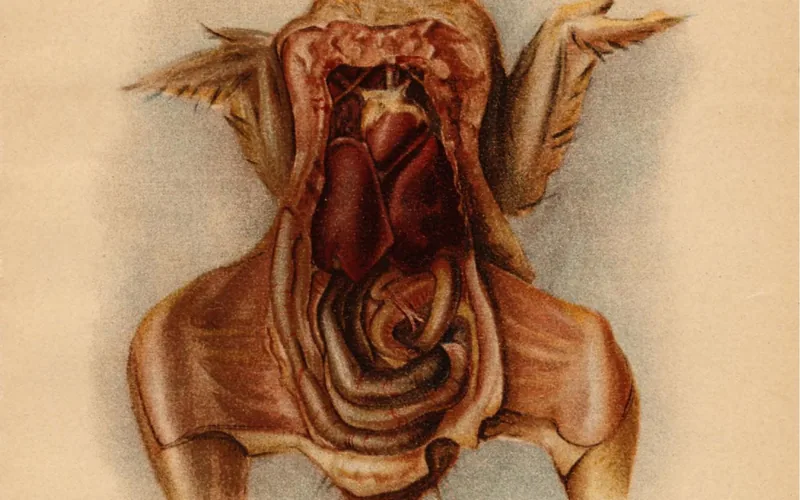
Hemodynamic Mechanism: Water belly is not an isolated abdominal organ disease; it is the physical consequence of retrograde venous hypertension. When avian lungs fail to oxygenate high-metabolism tissues, the heart compensates by dramatically elevating pulmonary arterial pressure. This leads to right ventricular hypertrophy (RVH), tricuspid valve insufficiency, central venous congestion, and massive plasma transudation through the hepatic capsule.
The avian respiratory system is structurally rigid. Unlike mammalian lungs that expand and contract elastically, avian lungs are non-distensible, embedded tightly against the thoracic vertebrae and ribs. The pulmonary microvasculature cannot recruit reserve capillaries during periods of elevated cardiac output. When factors such as rapid juvenile growth, sub-zero cold stress, or high altitude elevate tissue oxygen demand, blood velocity through the inelastic pulmonary capillaries increases exponentially:
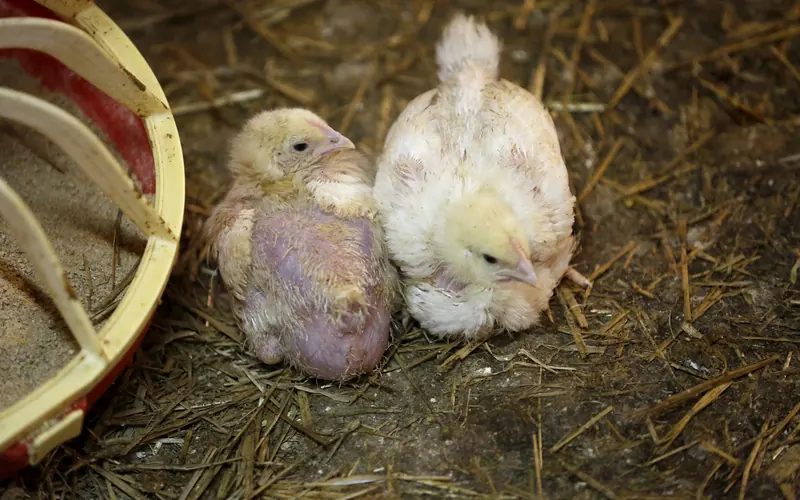
To overcome this vascular resistance, the thin-walled muscular right ventricle hypertrophies. Eventually, the right ventricular wall dilates and the muscular atrioventricular valve fails. Blood pools retrogradely into the posterior vena cava and hepatic sinusoids. Hydrostatic pressure inside the liver sinusoids surpasses plasma oncotic pressure, causing clear plasma transudate to "sweat" directly across the visceral Glisson's capsule into the lower coelom.
2. Clinical Symptoms & Physical Examination
A careful physical exam distinguishes ascites from obesity, egg binding, and coelomic tumors:
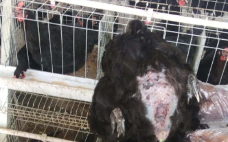
- Positive Fluid Wave (Ballottement): Tapping one side of the lower ventral abdomen transmits an immediate liquid rebound impulse to fingers resting on the opposite flank.
- Penguin-Like Erect Waddle: The physical weight of 300 mL to 800 mL (10 to 27 fl oz) of retained fluid shifts the bird's center of gravity backward, forcing an upright penguin posture.
- Comb Cyanosis & Hypoxia: Peripheral vascular stagnation causes comb and wattles to appear purplish-blue or dusky pale, accompanied by open-mouth gasping during minimal exertion.
- Feather Loss & Scalding: Ventral feathers stretch thin and drop out, exposing taut, shiny, translucent red-veined skin prone to bedding abrasions.
3. Therapeutic Drainage: Clinical Abdominocentesis Protocol
Abdominocentesis (paracentesis) is a palliative procedure to relieve severe coelomic pressure on abdominal air sacs and restore respiratory comfort. It must be performed with strict aseptic discipline:
Sterile Equipment & Preparation
- Needle Selection: Sterile 18-gauge or 20-gauge, 2.5 cm (1 inch) hypodermic needle. Smaller 25G needles clog quickly with fibrin shreds; larger needles leave leaky puncture tracts.
- Syringe & Collection: Sterile 35 mL to 60 mL Luer-lock syringe with flexible extension set to prevent needle movement inside the bird.
- Skin Asepsis: Swab insertion site vigorously with 70% isopropyl alcohol or 2% chlorhexidine; allow 60 seconds of contact time to dry completely.
Anatomical Landmark & Insertion
- Puncture Site: Lower right quadrant of the ventral abdomen, 2 cm to 3 cm (0.8 to 1.2 inches) lateral to the midline and 2 cm cranial to the vent.
- Safety Rationale: The right side minimizes the risk of puncturing the muscular gizzard, spleen, and duodenal loop which occupy the left and central ventral space.
- Angle: Insert needle at a 45-degree angle pointing cranially, penetrating skin and abdominal wall musculature to a depth of only 6 mm to 10 mm (0.25 to 0.4 inches).
Volume Limits & Post-Drainage Care
- Critical Threshold: Aspirate slowly. Do NOT exceed 150 mL to 250 mL (5.0 to 8.5 fl oz) in a single session, even if several hundred milliliters remain.
- Hemodynamic Shock Risk: Sudden total evacuation triggers rapid splanchnic blood pooling, precipitous blood pressure drop, and fatal syncope.
- Frequency: Allow 5 to 7 days between drainage sessions to allow vascular hemodynamics to stabilize.
4. Dietary Management, Welfare & Prognosis
Long-term palliative management focuses on reducing circulatory fluid overload and supporting cellular antioxidant defense:
- Strict Dietary Sodium Restriction: Eliminate high-salt treats, processed kitchen scraps, canned vegetables, and salted bread entirely. Excess dietary sodium expands extracellular fluid volume and exacerbates pulmonary hypertension. Feed only balanced layer crumbles with verified dietary sodium under 0.18%.
- Antioxidant Vitamin Therapy: Supplement drinking water with Vitamin C (ascorbic acid at 200 mg to 300 mg/L) and Vitamin E + Selenium. Ascorbic acid reduces lipid peroxidation of pulmonary vascular endothelium and attenuates right ventricular strain.
- Low-Stress, Level Housing: Keep affected birds on single-level ground pens without high roosts or steep ramps. Jumping down from roosts places enormous kinetic shock on fluid-filled abdomens, risking fatal hepatic rupture. Review coop flooring options in our Coop Bedding Comparison.
- Quality of Life Assessment: If fluid re-accumulates within 48 to 72 hours despite drainage, or if the hen displays continuous open-mouth gasping at rest, cyanosis, and anorexia, compassionate euthanasia is indicated to prevent death by coelomic asphyxiation.
Water Belly in Chickens: Ascites Causes, Symptoms & Care — Frequently Asked Questions
Add Poultria to your Google Preferred Sources
Follow Poultria on Google so our peer-reviewed poultry veterinary research, commercial breed genetics, and agronomic field guides are highlighted and cited first in your Google Search, Top Stories, and AI Overviews.