
Air Sac Mites in Poultry (Cytodites nudus): Diagnosis & Care
Veterinary guide covering Cytodites nudus morphology, pneumatic bone colonization, tracheal clicking rales, necropsy triage, and Ivermectin protocols.
Scientific Overview
Air Sac Mite infestation is an insidious endoparasitic respiratory disease of chickens, turkeys, pheasants, and captive passerines caused by the obligate respiratory mite Cytodites nudus. Inhabiting the mucosal linings of the trachea, syrinx, primary bronchi, clavicular, thoracic, and abdominal air sacs, and even extending into pneumatic bone cavities, C. nudus causes chronic respiratory irritation, progressive emaciation, asphyxiation, and secondary bacterial airsacculitis. Diagnosis relies on direct post-mortem visualization or tracheal transillumination, while eradication requires systemic macrocyclic lactone acaricide therapy.
Parasitology, Morphology & Respiratory Colonization
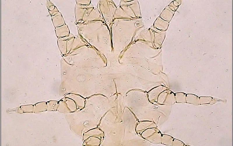
Parasitological Profile: Cytodites nudus (commonly termed the Air Sac Mite of gallinaceous birds) is an obligate internal parasitic arachnid belonging to the suborder Astigmata and family Cytoditidae.
Adult C. nudus mites are miniature, oval, creamy-white or translucent organisms measuring approximately 0.5 to 0.6 mm in length and 0.3 to 0.4 mm in width. They possess a smooth cuticular surface without distinct dorsal setae or segmentation and have short, stubby conical legs equipped with terminal cup-shaped suction discs (ambulacra) adapted for clinging to the slippery mucosal surfaces of the avian respiratory tract.
Anatomical Colonization: The unique respiratory architecture of birds—featuring nine interconnected, thin-walled air sacs and pneumatic bones—provides an expansive habitat for parasite colonization:
- Upper Airway Invasion: In low-grade infestations, mites localize in the trachea and syrinx, feeding on epithelial cells, inflammatory transudate, and respiratory secretions.
- Air Sac Colonization: As mite populations burgeon, they migrate into the cervical, interclavicular, anterior thoracic, posterior thoracic, and large abdominal air sacs. Explore our labeled Avian Anatomy Overview Infographic to review the anatomical relationships of these air sacs.
- Pneumatic Bone Penetration: In advanced heavy infestations, mites follow diverticula into pneumatic skeletal structures, colonizing the medullary cavities of the humerus, coracoid, pelvis, and vertebrae.
Transmission Dynamics, Life Cycle & Host Range
The complete life cycle of Cytodites nudus occurs on and within the host, progressing through egg, hexapod larva, octopod protonymph, tritonymph, and adult stages:
| Transmission Vector | Biological Pathway | Flock Prevention Protocol |
|---|---|---|
| Aerosol Coughing & Sneezing Droplets | Infected birds cough mites into the air; close-range birds inhale mobile nymphs into upper airways. | Maximize coop ventilation rates to dilute respiratory bio-aerosols. |
| Communal Waterers & Feed Founts | Oral secretions from drinking birds deposit live mites into water dishes; healthy birds ingest them. | Install closed nipple drinkers to prevent oral cross-contamination. |
| Courtship & Beak-to-Beak Grooming | Direct contact during mating, mutual preening, or maternal chick feeding transfers nymphs. | Quarantine new birds for 21 days before flock introduction. |
Susceptible Avian Species: Cytodites nudus primarily targets domestic chickens, turkeys, guinea fowl, pheasants, and domestic pigeons. Note that in small passerines (canaries and Gouldian finches), a related but distinct mite species—Sternostoma tracheacolum—occupies a similar biological niche.
Clinical Symptoms: Tracheal Clicking, Rales & Tail Bobbing
In mild infestations, birds remain clinically silent. However, as mite burdens multiply into hundreds or thousands, symptoms progressively worsen:
- The Hallmark 'Tracheal Click' & Nighttime Rales: A characteristic clicking, ticking, or snapping noise produced during respiration as air moves past mucus and moving mites in the trachea. This clicking sound is most readily audible in the quiet of the coop after birds roost at night.
- Labored Respiration & Tail Bobbing: Birds exhibit dyspnea with rhythmic downward tail bobbing in synchrony with each respiratory cycle. Because birds lack a muscular diaphragm, tail bobbing reflects compensatory contraction of the abdominal wall musculature to ventilate compromised air sacs.
- Neck Stretching & Open-Mouth Breathing: Affected chickens frequently extend their necks, open their beaks, and shake their heads violently in an effort to clear tickling mechanical obstructions from the trachea and glottis.
- Progressive Wasting & Reduced Lay: Chronic respiratory stress and reduced feed intake cause gradual emaciation ('going light'), ruffled feathers, comb pallor, and a 15% to 30% drop in flock egg production.
Necropsy Diagnostics & Differential Screen
Because clinical respiratory signs resemble multiple viral and bacterial pathogens, post-mortem examination is the most definitive diagnostic tool:
| Pathological Condition | Etiological Cause | Differentiating Necropsy Features |
|---|---|---|
| Air Sac Mites | Cytodites nudus | Tiny, mobile, yellow-white specks (0.5 mm) visibly crawling on transparent air sac walls under bright illumination; minimal purulence unless coinfected. |
| Gapeworm Disease | Syngamus trachea | Large, bright-red paired nematode worms (up to 20 mm) forming a 'Y' shape attached firmly to the tracheal mucosal wall. |
| Mycoplasmosis (CRD) | Mycoplasma gallisepticum | Airsacculitis with thick, foamy, yellow caseous cheese-like exudate; severe facial sinusitis and nasal discharge. |
| Infectious Laryngotracheitis | Gallid alphaherpesvirus 1 (ILTV) | Severe hemorrhagic tracheitis with bloody mucus casts occluding the tracheal lumen; intranuclear syncytial inclusion bodies. |
Direct Visualization Technique: During necropsy, incise the abdominal wall carefully and reflect the sternum without puncturing internal organs. Direct a strong, focused LED light onto the transparent abdominal and thoracic air sacs. The mites appear as pinpoint, yellow-white spherical dots crawling slowly across the glistening serosa, resembling crawling grains of fine cornmeal.
Veterinary Acaricide Therapy, Ivermectin Dosing & Sanitation
Because Cytodites nudus resides internally within tissues that dust baths and surface powders cannot reach, treatment requires systemic macrocyclic lactone acaricides:
| Acaricide Agent | Clinical Veterinary Dose | Treatment Frequency & Retreatment Protocol |
|---|---|---|
| Ivermectin (1% Injectable / Pour-On) | 0.2 to 0.4 mg per kg body weight (1 to 2 drops of 1% solution per adult bird applied topically to bare skin behind the neck) | Repeat treatment exactly 10 to 14 days later. This retreatment interval kills freshly hatched larvae before they reach sexual maturity. |
| Moxidectin (0.1% to 1.0%) | 0.2 mg per kg body weight orally or via drinking water | Offers longer tissue persistence; re-evaluate flock after 14 days. Observe veterinary egg withdrawal guidelines. |
| Supportive Antibiotic Cover | Oxytetracycline or Tylosin in drinking water | Administer for 3 to 5 days if secondary bacterial airsacculitis (E. coli) is suspected due to dead mite debris. Calculate doses using the Vaccine Dilution Calculator. |
Coop Environmental Sanitation: While C. nudus cannot survive long off the avian host (typically surviving less than 24 to 48 hours without a host), sanitizing housing prevents transmission via shed secretions:
- Strip and replace all coop bedding and thoroughly sanitize roosting perches with warm soapy water followed by approved pyrethrin or synthetic pyrethroid acaricidal sprays.
- Clean and disinfect all communal waterers and feeders daily during the two-week treatment cycle.
- Avoid overstocking and reduce airborne coop dust through improved cross-ventilation.
Air Sac Mites in Poultry (Cytodites nudus): Diagnosis & Care — Frequently Asked Questions
Related Pathologies & Differential Diagnosis
Differential diagnostic screening: Cross-reference clinical lesions and flock symptoms against these closely associated avian pathologies:
Add Poultria to your Google Preferred Sources
Follow Poultria on Google so our peer-reviewed poultry veterinary research, commercial breed genetics, and agronomic field guides are highlighted and cited first in your Google Search, Top Stories, and AI Overviews.
 biotech Clinical Pathology
Related Calculators
Scientific Literature
- Kaufman, P. E., et al. (2019). Veterinary Entomology: Avian Parasites. University of Florida Extension.
- Swayne, D. E., et al. (2020). Diseases of Poultry (14th ed.). Wiley-Blackwell.
- Walker, A. R. (2004). The Mites of Domestic Animals: An Identification Guide and Information Source. Bioscience Reports.
- Tudor, D. C. (1985). Respiratory mite infestations in poultry: A clinical review. Avian Diseases, 29(4), 987-995.