
Blood Spots vs Meat Spots in Eggs: Causes & Safety Guide
Differentiating follicular capillary hemorrhage from oviduct mucosal inclusions: biological triggers, commercial grading tolerances, and flock triage.
Scientific Overview
Cracking an egg into a morning skillet only to spot a bright red droplet or a tiny brownish fleck on the yolk is an unsettling surprise for any cook. If you are asking blood spot in egg is it safe to eat or wondering about blood spot in egg yolk causes, rest assured that this is a completely natural biological occurrence, not a fertilized chick or an infection. Known clinically as blood spots and meat spots, these inclusions happen when a microscopic capillary breaks as the yolk releases from the hen's ovary, or when a tiny particle of oviduct lining sloughs off during albumen formation. This veterinary guide explains the biological difference between blood spots and meat spots, clarifies USDA grading standards, and confirms that eggs with small spots are 100% safe to eat when cooked to 165°F (74°C).
1. Ovarian & Oviducal Biology: Follicular Stigma Rupture vs Tissue Sloughing
Biological Origin: In laying hens, ovulation occurs approximately 30 to 45 minutes after the previous egg is laid. The mature ovarian follicle is wrapped in an extensive capillary network except along a narrow, avascular line called the stigma or suture line. Under normal conditions, the follicle splits cleanly along this avascular seam to release the yolk. A blood spot occurs when an aberrant micro-capillary crosses the stigma and ruptures during ovulatory tear.
To understand the true nature of internal egg inclusions, we must distinguish between where in the reproductive tract the defect originates:
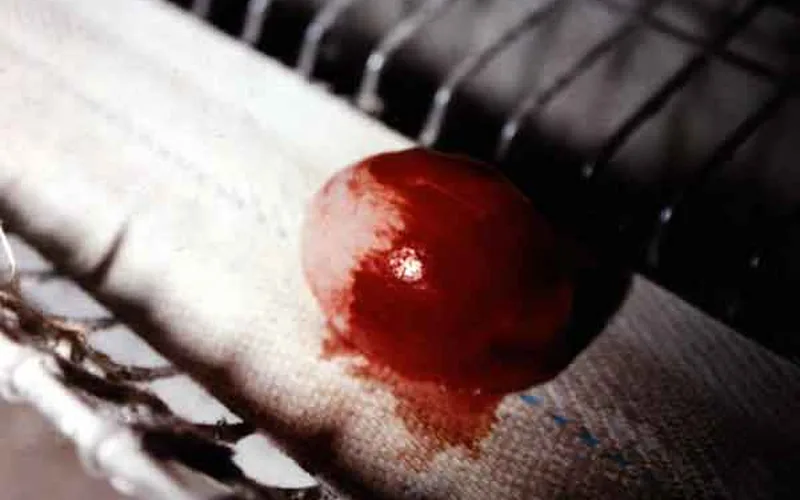
Blood Spot Anatomy (Ovary)
- Location: Directly attached to the yolk's vitelline membrane surface, or rarely free in the albumen.
- Appearance: Bright red, scarlet, or ruby drop. If the egg is stored for several weeks, the hemoglobin oxidizes to dull brown.
- Mechanism: Follicular capillary cross-rupture during ovulation. If bleeding is extensive, blood diffuses throughout the albumen, producing a "bloody egg" (bloody white).
Meat Spot Anatomy (Oviduct)
- Location: Suspended inside the thick or thin albumen, entirely separate from the yolk.
- Appearance: Fleck or ribbon colored brownish-tan, grey, or off-white; firm rubbery texture.
- Mechanism: Cellular debris sloughed from the mucosal lining of the oviduct magnum or isthmus, or an old ovarian blood clot that underwent chemical transformation and bleaching in the acidic albumen.
For background on how egg freshness and albumen density are measured scientifically, read our guide on the Haugh Unit egg quality benchmark.
2. USDA & International Commercial Grading Standards
The United States Department of Agriculture (USDA) Agricultural Marketing Service and the European Commission establish stringent legal classification tolerances for blood and meat spots in commercial table eggs:
| Commercial Grade | Allowable Spot Diameter | Location & Type Tolerance | Market Destination |
|---|---|---|---|
| Grade AA | Zero prominent spots allowed | Completely free from visible blood or meat inclusions. | Premium retail table carton eggs. |
| Grade A | Zero prominent spots allowed | Albumen must be practically free of foreign material. | Standard grocery retail cartons. |
| Grade B | Aggregate diameter < 3.0 mm (1/8 in) | Small blood or meat spots confined to yolk surface or white. | Institutional baking, commercial liquid egg breakers. |
| Inedible / Loss | Aggregate diameter ≥ 3.0 mm (1/8 in) | Large bloody inclusions, blood diffused throughout white, rot. | Condemned from human food supply; animal feed processing. |
In commercial packing stations, electronic spectrophotometric candling scanners process over 150,000 eggs per hour. These cameras transmit specific wavelengths through the shell; hemoglobin absorbs light at 577 nm, allowing robotic arms to automatically eject eggs containing blood spots over 3 mm.
3. Interactive Tool: Egg Grade & Defect Triage Tool
Use this diagnostic triage tool to evaluate an cracked egg's defect type, size, and shell characteristics to determine its commercial grade and culinary safety:
🍳 Egg Grade & Defect Triage Tool
USDA/EU Quality Standard4. Flock Diagnostic Checklist: Genetics, Vitamins A & K, and Mycotoxins
While occasional blood spots (under 2% to 4% of total eggs in brown flocks) are completely normal, a sudden spike where 10% to 25% of eggs contain blood or meat spots indicates flock-wide physiological distress. Use this clinical checklist to investigate:
- Vitamin A Hypovitaminosis: Vitamin A maintains the structural integrity of stratified squamous and columnar epithelial membranes. When dietary Vitamin A drops below 6,000 IU/kg, ovarian follicular capillaries become abnormally fragile. Inspect feed manufacturing dates; Vitamin A oxidizes rapidly in stale feed stored over 60 days in warm climates. Supplement feed with stabilized synthetic retinyl acetate.
- Vitamin K Deficiency & Antagonists: Vitamin K is the co-factor for gamma-glutamyl carboxylase, synthesizing prothrombin and blood clotting factors VII, IX, and X. If layers consume feeds contaminated with sulfa medications (like sulfamethazine) or anticoagulant rodenticides (warfarin or brodifacoum), clotting time lengthens dramatically. What would have been an invisible micro-bleed expands into a major bloody white.
- Mycotoxicosis (T-2 Toxin & Ochratoxin A): Trichothecene mycotoxins from moldy corn or wheat cause vascular necrosis, capillary fragility, and oral lesions. Test grains for mold contamination and introduce high-capacity aluminosilicate or yeast cell wall mycotoxin binders into the layer mash.
- Acoustic & Predator Disturbance: Acute adrenaline release stimulates involuntary oviduct contractions and sudden blood pressure spikes. Loose barking dogs, nocturnal rodent infestations, or malfunctioning fan belts that screech suddenly can double the incidence of blood spots within 48 hours.
If you also observe oviduct inflammation, yellow caseous exudate, or abdominal swelling alongside internal inclusions, evaluate your birds for salpingitis and egg yolk peritonitis.
Blood Spots vs Meat Spots in Eggs: Causes & Safety Guide — Frequently Asked Questions
Add Poultria to your Google Preferred Sources
Follow Poultria on Google so our poultry veterinary references, commercial breed genetics, and agronomic field guides are highlighted and cited first in your Google Search, Top Stories, and AI Overviews.