Egg Bound Chicken Symptoms: Emergency Care & Calcium Guide
Clinical veterinary triage for oviduct impaction: recognizing early warning signs, calcium gluconate administration, warm hydrotherapy, and life-saving cloacal delivery protocols.
Scientific Overview
Egg binding (dystocia) is a critical avian reproductive emergency in which a calcified or soft-shelled egg becomes physically lodged in the hen's oviduct or cloaca. In modern backyard flocks—particularly high-producing production hybrids and aging heritage layers—egg binding can progress from mild discomfort to fatal cloacal necrosis, oviduct rupture, or egg yolk peritonitis within 24 to 48 hours. Because avian kidneys sit directly against the synsacrum adjacent to the pelvic canal, a lodged egg places continuous mechanical pressure on renal blood vessels and the sciatic nerve plexus, inducing rapid hypovolemic shock and bilateral hindlimb paresis. Immediate clinical intervention is essential to relieve impaction and stabilize the hen.
1. Clinical Pathophysiology: Why Egg Binding Kills Rapidly
Emergency Triage Alert: Egg binding is a life-threatening physiological blockage, not a passive resting phase. When an egg lodges in the caudal magnum, isthmus, uterus (shell gland), or vagina, normal cloacal defecation is obstructed. The mass compresses the external iliac veins and pelvic nerves, leading to leg paralysis, toxic urate retention, and circulatory collapse within 24 to 48 hours.
Avian egg development is an extraordinary 25-to-26-hour sequence. Every egg requires the deposition of approximately 2.0 g to 2.3 g (0.07 to 0.08 oz) of pure calcium carbonate onto the organic shell matrix inside the uterus. When a hen's dietary calcium intake is inadequate or medullary bone reserves become exhausted, circulating ionized calcium (iCa) drops precipitously. Because smooth muscle contractions depend directly on extracellular calcium influx, the muscular uterine wall (myometrium) enters hypocalcemic tetany or atony—losing the contractile force required to push the egg past the muscular vaginal sphincter into the cloaca.
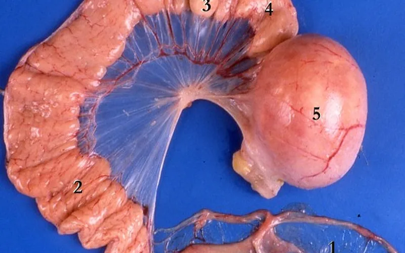
Compounding the muscular failure, the avian pelvic inlet is bounded dorsally by the rigid fused synsacrum. Unlike mammals whose pelvic ligaments soften significantly under relaxin hormone surges, the avian pelvic girdle cannot expand. If an egg is abnormally large, double-yolked, rough-shelled, or misaligned horizontally, it acts as a mechanical wedge. Compression of the adjacent renal portal venous system halts normal urinary filtration, causing rapid hyperuricemia, dehydration, and systemic shock.
2. Symptom Recognition & Differential Diagnosis
Accurate diagnosis begins with distinguishing dystocia from common conditions that mimic nesting behavior or abdominal distress, such as broodiness, coccidiosis, and water belly:
| Clinical Indicator | Egg Bound (Dystocia) | Broody Hen | Ascites (Water Belly) |
|---|---|---|---|
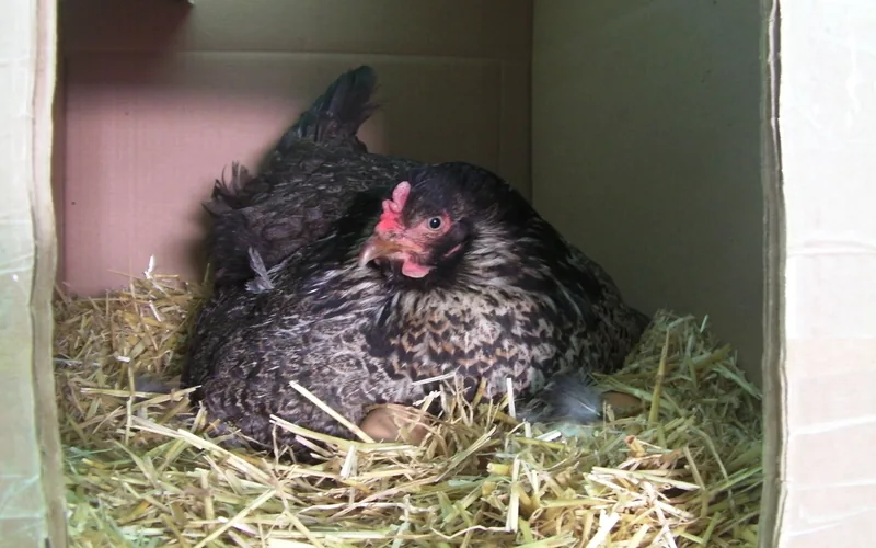
| Physical Posture | "Penguin stance" (upright torso), wide-legged stance, tail pumping up and down rhythmically. | Flattened "pancake" posture in nest box, fluffed neck hackles, protective growling. | Upright waddle, fluid-distended lower abdomen, resting frequently on keel bone. |
| Abdominal Palpation | Firm, hard, distinct oval mass palpable between pubic pin bones (or cranial to vent). | Soft, pliable abdomen, warm featherless brood patch on breast. | Fluctuant, squishy, fluid-wave feeling; no distinct solid calcified shell. |
| Defecation Patterns | Complete cessation of droppings or small, watery white urate smears with fecal straining. | Infrequent, massive, pungent "broody droppings" when leaving nest box briefly. | Normal to loose watery droppings; appetite remains moderate until end stage. |
| Locomotion & Reflexes | Unsteady gait, frequent collapsing, unilateral or bilateral leg paralysis from sciatic pinch. | Fully mobile, brisk defensive pecking, walks normally when removed from nest. | Reluctant to walk long distances due to weight; no sciatic nerve paralysis. |
3. Immediate 4-Step Medical Protocol for Egg-Bound Hens
Follow this sequential, clinically validated protocol immediately upon identifying an egg-bound hen. Time is the single greatest determinant of survival:
Step 1: Emergency Calcium Administration
Smooth muscle exhaustion cannot be resolved by heat alone. Administer bioavailable calcium directly to restore myometrial contractility:
- Liquid Calcium Gluconate (10%): Administer 1.0 mL per kg (0.45 mL per lb) of body weight orally or via subcutaneous injection in the inguinal fold.
- Over-the-Counter Alternative: A crushed 500 mg to 600 mg calcium carbonate + Vitamin D3 tablet dissolved in 3 mL of warm water given slowly via oral syringe.
- Action: Rapidly spikes serum ionized calcium, triggering strong, rhythmic expulsive uterine contractions within 30 to 60 minutes.
Step 2: Warm Hydrotherapy (Epsom Bath)
Submerge the lower half of the hen's body in a warm shallow bath to relax pelvic musculature:
- Water Temperature: Calibrate precisely to 38.5°C to 40.0°C (101°F to 104°F). Never exceed 41°C (106°F) to avoid hyperthermia.
- Mineral Additive: Dissolve 50 g (1.8 oz / roughly 1/4 cup) of magnesium sulfate (Epsom salt) per 4 liters (1 gallon) of water to ease muscle spasms.
- Duration & Safety: Soak for 20 minutes. Support the bird continuously; an exhausted hen can easily slip underwater and drown.
Step 3: Cloacal Lubrication & Thermal Chamber
Reduce frictional resistance and provide an optimal metabolic microclimate:
- Lubricant: Gently infuse 2 mL to 3 mL of sterile, water-soluble surgical lubricant (K-Y Jelly) 1 cm to 2 cm (0.5 to 0.8 inches) inside the vent using a blunt, needleless syringe.
- Recovery Chamber: Place the hen in a warm, dark, humid nursery box maintained at 28°C to 30°C (82°F to 86°F) with 70%+ relative humidity (using a warm, damp towel beneath bedding).
- Rest: Leave the bird undisturbed in total darkness for 2 hours. Over 80% of uncomplicated cases deliver the egg naturally during this resting window.
Step 4: Surgical Veterinary Intervention
If the egg remains unpassed after 4 to 6 hours or the hen exhibits open-mouth breathing:
- Veterinary Consultation: Seek immediate avian veterinary care for digital manipulation under sedation or prostaglandin E2 (PGE2) gel cloacal priming.
- Controlled Ovocentesis: A veterinarian may insert an 18-gauge needle through the dilated vent into the exposed egg pole, aspirate albumen and yolk, and gently collapse the shell inward.
- Prognosis: Prolonged impactions exceeding 24 hours carry significant risks of salpingitis, peritonitis, and permanent oviduct scarring.
4. Post-Delivery Recovery & Preventing Recurrence
Passing a retained egg is an exhausting trauma that leaves the cloacal and uterine tissues inflamed, bruised, and susceptible to secondary infection or prolapse:
- Electrolyte & Carbohydrate Replenishment: Offer cool drinking water infused with poultry electrolytes and 1% glucose or dextrose to reverse dehydration and lactic acid buildup from prolonged muscular straining.
- Temporary Ovulation Suppression: To give an inflamed oviduct time to heal, reduce photoperiod exposure to 8 to 10 hours of dim light for 5 to 7 days. This temporarily pauses pituitary LH surges and halts ovulation.
- Coarse Oyster Shell Kinetic Balance: Ensure free-choice coarse oyster shell (particle size 2 mm to 5 mm) is always provided in separate hoppers. Coarse calcium dissolves slowly in the gizzard over 16 to 20 hours, supplying circulating calcium during the dark night hours when eggshell calcification occurs. For precise feeding guidelines, read our Laying Hen Feed Guide: 16%–18% Protein Rations & Calcium Curves.
- Body Condition Scoring: Eliminate high-fat treats, excess corn, and table scraps that cause obesity. Fat hens deposit adipose tissue around the pelvic canal, mechanically restricting oviduct dilation. Check optimal breed body weights in our Best Egg Laying Chickens Comparison.
Egg Bound Chicken Symptoms: Emergency Care & Calcium Guide — Frequently Asked Questions
Add Poultria to your Google Preferred Sources
Follow Poultria on Google so our peer-reviewed poultry veterinary research, commercial breed genetics, and agronomic field guides are highlighted and cited first in your Google Search, Top Stories, and AI Overviews.