
Chicken Diarrhea: Causes, Coccidiosis & Dropping Colors Triage
Veterinary clinical triage of watery droppings: differentiating normal cecal evacuations from bloody coccidiosis, green starvation bile, yellow enteritis, and white renal urates.
Scientific Overview
Abnormal droppings are frequently the earliest visible indicator of systemic distress, metabolic imbalance, or gastrointestinal pathogen proliferation in poultry. Because birds possess a cloaca—a single shared exit chamber for fecal material, urinary urates, and reproductive output—evaluating chicken diarrhea requires distinguishing true enteric enteritis from normal physiological variations. While a normal hen produces firm, brown digestive droppings capped with creamy white uric acid crystals 12 to 16 times daily alongside pasty caramel-colored cecal droppings, watery or discolored feces demand immediate diagnostic triage. Whether managing commercial broiler hybrids like Ross 308 and Cobb 500 where rapid transit and wet litter threaten flock footpad health, or a heritage laying flock, this guide details clinical differential diagnosis, anticoccidial dosing, and gut microbiome recovery.
1. Fecal Physiology: Normal Enteric vs. Cecal Droppings vs. Diarrhea
Avian Physiological Fact: Because chickens lack a urinary bladder and urethra, the kidneys excrete nitrogenous waste as insoluble white uric acid crystals (urates) directly through the ureters into the urodeum of the cloaca. A normal healthy dropping consists of a firm, coiled core of digested plant chyme topped with a distinct white uric acid cap.
In modern poultry husbandry, evaluating droppings is a core clinical skill. Highly productive meat birds like Ross 308 broilers consume up to double their body weight in water relative to feed. Their high metabolic throughput produces voluminous, moist droppings that can easily be mistaken for pathological enteritis if the keeper does not understand gut transit physiology.
Normal Intestinal Droppings
Passed 12 to 16 times daily. Cylindrical, semi-solid, brown, greenish-brown, or greyish. Coated with clean white urate caps. Minimal odor. Indicates balanced water-to-feed intake and intact intestinal villi.
Normal Cecal Droppings
Passed 2 to 3 times daily. Pasty, unformed, sticky, caramel-to-dark-brown. Has a distinct pungent fermented smell. Result of anaerobic microbial digestion of complex polysaccharides in the blind ceca. Frequently misidentified as diarrhea.
2. Clinical Triage: Dropping Color Diagnostics Matrix
When true diarrhea occurs, the color, consistency, and accompanying systemic symptoms pinpoint the underlying etiology. Use the clinical matrix below for rapid flock triage:
| Dropping Appearance | Primary Differential | Pathological Mechanism | Action Protocol |
|---|---|---|---|
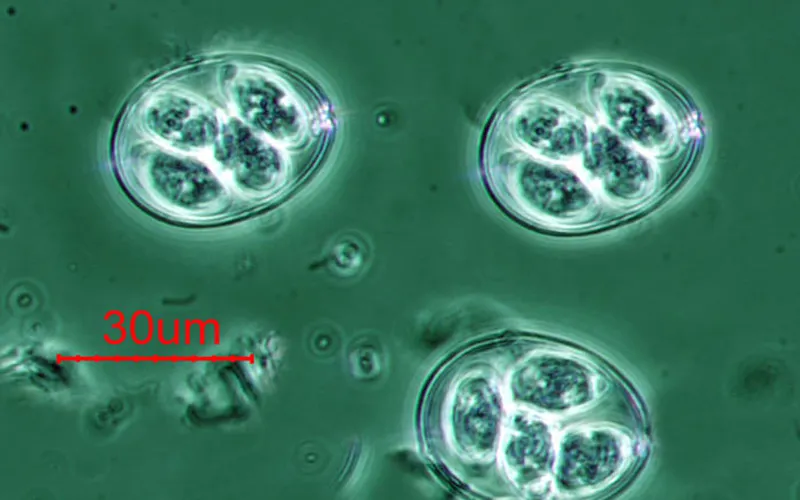
| Bright Red or Frank Blood | Cecal Coccidiosis (Eimeria tenella) |
Second-generation schizonts rupture cecal capillaries, causing acute luminal hemorrhage and hypovolemia. | Immediate Amprolium (Corid) in water; warm brooder; vitamin K injection if prescribed. |
| Bright Neon Green | Anorexia / Starvation Bile (Or Marek's / ND / Lead) |
Bile (biliverdin) continues secreting into empty gut. Concentrated, unabsorbed green pigments pass into cloaca. | Check crop for sour or impacted crop; evaluate for Marek's paralysis; tube-feed electrolyte-glucose slurry. |
| Sulfur-Yellow / Foamy | Blackhead (Histomoniasis) (Or infectious coryza / typhoid) |
Histomonas meleagridis necrotizes cecal mucosa and invades liver parenchyma, discharging yellow necrotic casts. | Isolate immediately; keep away from turkeys; veterinary dimetridazole/metronidazole where legal. Read our Blackhead Disease Guide. |
| Chalky White Watery | Vent Gleet or Renal Failure (Urate nephrosis / IBV) |
Candidiasis cloacitis or infectious bronchitis nephropathic strains causing massive excess uric acid discharge. | Isolate, bathe vent in warm chlorhexidine, oral nystatin or fluconazole. Read our Vent Gleet Guide. |
| Clear Copious Water | Heat Stress Polydipsia (Excessive summer drinking) |
Over-consumption of water to dissipate body heat flushes straight through the kidneys and cloaca. | Provide shade, cool fresh water, electrolytes, and evaporative cooling. Review Heat Stress Cooling Protocols. |
3. Interactive Fecal Triage & Clinical Assessment Tool
Avian Fecal Symptom & Pathology Matcher
Select the observed dropping characteristics and secondary flock symptoms below to identify the most probable clinical cause and veterinary recommended intervention:
Rupture of intestinal capillary plexus caused by actively replicating coccidial schizonts. High risk of dehydration and acute flock mortality within 48 hours.
4. Treatment Protocols: Coccidiostats, Electrolytes & Gut Recovery
Managing an outbreak of poultry diarrhea requires a sequential three-phase therapeutic approach: halting pathogen replication, rehydrating the hypovolemic host, and restoring the epithelial mucosal barrier.
- Therapeutic Coccidiostat Administration: For coccidiosis, start Amprolium (Corid) at 9.5 mL per 3.8 L (2 tsp/gal) for 5 days, or Toltrazuril (Baycox 2.5%) at 7 mg/kg body weight daily for 2 consecutive days via drinking water. Toltrazuril acts on all intracellular developmental stages of Eimeria, providing rapid clinical arrest.
- Balanced Osmotic Rehydration: Severe watery diarrhea flushes essential sodium, potassium, and bicarbonate ions from systemic circulation, triggering metabolic acidosis. Dissolve a balanced poultry electrolyte pack into drinking water, or formulate an emergency oral rehydration solution: 4.5 g (1 tsp) salt, 2.5 g (0.5 tsp) baking soda, and 20 g (1.5 tbsp) dextrose or sugar per 1 Liter (1.05 qt) of water.
- Microbiome Competitive Exclusion: Following pharmaceutical anticoccidials or antibiotics, the denuded intestinal lumen is highly vulnerable to colonization by pathogenic Clostridium perfringens, leading to Necrotic Enteritis. Seed the gut with beneficial Lactobacillus acidophilus, Bacillus subtilis, and organic yeast cultures (MOS) to re-establish mucosal competitive exclusion.
- Litter Moisture Remediation: Diarrhea elevates house moisture, causing litter caking and ammonia volatilization. To prevent footpad dermatitis and manage moisture, review our Litter Moisture Calculator and Minimum Ventilation Guide.
Chicken Diarrhea: Causes, Coccidiosis & Dropping Colors Triage — Frequently Asked Questions
Add Poultria to your Google Preferred Sources
Follow Poultria on Google so our peer-reviewed poultry veterinary research, commercial breed genetics, and agronomic field guides are highlighted and cited first in your Google Search, Top Stories, and AI Overviews.