
Avian Coccidiosis: Symptoms, Lesions & Treatment Guide
A clinical field manual covering 7 Eimeria species, Johnson & Reid lesion scoring, and Amprolium protocols.
Scientific Overview
Avian coccidiosis is an acute or subclinical intestinal infection caused by protozoan parasites of the genus Eimeria. It represents the single most costly parasitic challenge in broiler and layer farming worldwide, causing suppressed growth, poor Feed Conversion Ratio (FCR), mucosal sloughing, and flock mortality. Detecting coccidiosis in chickens symptoms early is critical: once clinical bloody droppings or severe depression appear in a poultry house, subclinical intestinal damage has already compromised nutrient absorption across hundreds of pen mates.
1. Etiology & Life Cycle: Understanding the Eimeria Parasite
Coccidiosis is not caused by bacteria or viruses, but by microscopic single-celled protozoa. While domestic fowl can host up to nine identified species, seven cause significant economic disease: Eimeria acervulina, Eimeria maxima, Eimeria tenella, Eimeria brunetti, Eimeria necatrix, Eimeria mitis, and Eimeria praecox. Each species exhibits strict site-specificity, invading distinct anatomical zones of the digestive tract.
The parasite multiplies through an obligate direct life cycle lasting 4 to 7 days:
- Exogenous Sporulation: Non-infectious unsporulated oocysts are passed in chicken droppings. In warm, moist litter at 25.0°C to 30.0°C (77.0°F to 86.0°F) with relative humidity above 60%, the oocyst sporulates within 24 to 48 hours, forming four sporocysts containing two infective sporozoites each.
- Mechanical Excystation: When ingested, the gizzard's muscular grinding and bile salts in the duodenum break open the oocyst shell, liberating active sporozoites.
- Schizogony (Asexual Cycling): Sporozoites penetrate gut epithelial enterocytes, dividing rapidly into thousands of merozoites across 2 to 4 asexual generations, rupturing host intestinal cells.
- Gametogony (Sexual Reproduction): Merozoites differentiate into microgametocytes (male) and macrogametocytes (female). Fertilization produces fresh unsporulated oocysts shed back into the bedding.
2. Clinical Symptoms: Identifying Coccidiosis in Chickens
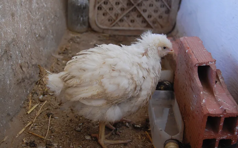
Recognizing coccidiosis in chickens symptoms and early coccidia symptoms in chickens prevents minor digestive upsets from escalating into whole-house mortality events. Typical signs depend on the bird's age, immune readiness, and the infecting Eimeria species:
- Frank red blood in droppings or reddish-black tarry stools (classic E. tenella).
- Severe dehydration with shriveled, pale combs and pale leg shanks.
- Huddling under brooder heaters with ruffled feathers and closed eyes.
- Complete feed refusal (anorexia) paired with elevated water consumption.
- Orange mucus and undigested feed grain passed in moist droppings.
- Uneven flock uniformity; stunted runts mixed among healthy birds.
- Elevated Feed Conversion Ratio (FCR) rising by 0.05 to 0.15 points.
- Poor pigment absorption causing pale egg yolks and yellow shank fade.
Chicks are most susceptible between 3 and 6 weeks of age, when maternal egg-yolk antibodies have fully waned but active cellular gut immunity is still developing. In backyard pullets, outbreaks frequently follow rainy spells or wet bedding around leaky nipple drinkers.
3. Anatomical Lesion Profiling & Johnson & Reid (0–4) Scoring
Accurate species diagnosis requires field post-mortem necropsy. Poultry veterinarians utilize the standardized Johnson & Reid (1970) Lesion Scoring Index (graded 0 for normal gut tissue up to +4 for fatal necrosis):
| Eimeria Species | Primary Gut Location | Pathognomonic Gross Lesions | Severity Index |
|---|---|---|---|
| Eimeria tenella | Cecal Pouches | Distended, ballooned ceca filled with frank clotted blood, core caseous plugs, sloughed mucosa | High (Up to +4) |
| Eimeria acervulina | Duodenal Loop | White transverse ladder-like stripes or oval plaques arranged across mucosal folds | Moderate (+1 to +3) |
| Eimeria maxima | Mid-Gut (Jejunum) | Thickened gut wall, orange mucus, pinpoint petechiae; primary trigger for Necrotic Enteritis | Severe (+2 to +4) |
| Eimeria necatrix | Mid-Gut & Ceca | Severe ballooning of mid-small intestine with 'salt-and-pepper' white and red necrotic foci | Fatal (+4) |
| Eimeria brunetti | Lower Intestine & Rectum | Coagulative necrosis, catarrhal enteritis, cheese-like mucosal slough near cloaca | Moderate (+2 to +3) |
4. Veterinary Treatment Protocols: Medication & Dosing
Once clinical disease breaks out, feed-grade coccidiostats are ineffective because sick birds stop eating but continue drinking. Immediate drinking water medication is mandatory:
- Amprolium Therapy: The most common field medication. Administer Amprolium 9.6% oral solution at 240 mg per liter (908 mg per gallon) of drinking water (equivalent to 2.5 mL per liter / 9.5 mL per gallon) continuously for 5 consecutive days, followed by a reduced maintenance dose of 60 mg per liter (227 mg per gallon) for 7 additional days. Amprolium acts as a thiamine (Vitamin B1) competitor, starving developing merozoites. Do not administer supplemental Vitamin B1 during treatment.
- Toltrazuril (Baycox): Highly effective symmetrical triazinone. Administer at 25 ppm (7 mg per kg body weight / 3.2 mg per lb) continuously in drinking water for 2 consecutive days, or at 75 ppm for 8 hours daily over 2 days. Toltrazuril acts on all intracellular developmental stages without compromising natural immunity development.
- Sulfadimethoxine / Trimethoprim-Sulfa: Broad-spectrum sulfonamide administered at 0.05% in drinking water for 6 days. Particularly valuable when secondary bacterial enteritis (Clostridium perfringens) complicates mucosal bleeding.
Following anticoccidial treatment, administer water-soluble Vitamin A and Vitamin K3 (menadione) at 5 mg per liter (19 mg per gallon) to accelerate intestinal mucosal healing and clotting factor synthesis.
5. Prevention Programs: Vaccines, Shuttle Programs & Sanitation
Long-term control relies on proactive biosecurity and controlled immune exposure rather than continuous medication:
Live Coccidiosis Vaccines
Live attenuated or non-attenuated vaccines (e.g., Paracox, Coccivac, Immucox) are sprayed onto day-old chicks in hatchery cabinets using an edible dye. Chicks preen the droplet-delivered oocysts from down feathers. Ingested parasites cycle through the gut over 3 successive generations in the brooder house, building life-long protective immunity.
Chemical & Ionophore Shuttle Programs
To prevent drug resistance, commercial integrators rotate synthetic chemicals (Nicarbazin, Diclazuril, Robenidine) in starter rations, transitioning to polyether ionophores (Salinomycin, Monensin, Lasalocid) in grower diets. Shuttle programs prevent continuous selective pressure on wild parasite populations.
Keep litter moisture strictly below 25% by running adequate minimum ventilation fans. Eimeria oocysts cannot sporulate without free moisture; dry bedding breaks the transmission chain before clinical disease manifests.
 biotech Clinical Pathology
Related Calculators
Scientific Literature
- Johnson, J., & Reid, W. M. (1970). Anticoccidial drugs: lesion scoring techniques in battery and floor-pen experiments with chickens. Experimental Parasitology, 28(1), 30-36.
- Diseases of Poultry, 14th Edition (David E. Swayne et al., 2020).
- World Organisation for Animal Health (WOAH). Manual of Diagnostic Tests and Vaccines for Terrestrial Animals.