
Chicken Gross Necropsy Lesion Decision Tree
Interactive post-mortem decision tree and avian veterinary differential diagnosis engine. Record observed macroscopic organ lesions across 8 anatomical systems, examine 35 verified reference necropsy photographs, differentiate between statutory viral emergencies (HPAI, Velogenic Newcastle) and endemic bacterial/protozoal flock infections, and generate a printable veterinary post-mortem field record.
grid_view Systematic Organ-by-Organ Post-Mortem Lesion Index
Evaluate poultry mortalities across 8 physiological systems covering 65 macroscopic lesion indicators calibrated to AAAP necropsy diagnostic standards:
Comb, wattles, infraorbital sinuses, conjunctiva, and oral cavity
- Dark cyanosis & severe edema
- Facial edema & infraorbital swelling
- Distended with caseous cheesy pus
- Copious putrid foul nasal discharge
- + 6 more lesion markers
Tracheal lumen, bronchi, lung parenchyma, and 9 air sacs
- Frank blood clots & hemorrhagic casts
- Mucosal petechiae & congestion
- Clear to catarrhal serous mucus
- Catarrhal-hemorrhagic exudate
- + 6 more lesion markers
Ingluvial crop mucosa, glandular stomach, and muscular gizzard
- Petechiae on mucosal papillae tips
- Hemorrhages at proventricular junction
- Thickened white curd-like pseudomembrane ('thrush')
- Severe koilin ulceration & sloughing
Duodenal loop, jejunum, ileum, cecal tonsils, and blind ceca
- Severely ballooned with dark blood & clots
- Firm laminated yellow-green caseous cores
- Swollen, dark hemorrhagic patches
- Friable tan 'Turkish towel' necrotic membrane
- + 5 more lesion markers
Hepatobiliary parenchyma, reticuloendothelial spleen, and heart sac
- Circular depressed 'bullseye' necrotic craters
- Pinpoint white necrotic spots ('starry sky')
- Thick white-yellow fibrinous capsule ('frosted')
- Fibrinous pericardial sac exudate & adhesion
- + 7 more lesion markers
Primary lymphoid tissues regulating humoral and cellular immunity
- Swollen (2-3x), edematous, yellow gelatinous transudate
- Severe intra-bursal blood clots & mucosal petechiae
- Severe atrophy (< 1/3 normal size) & depletion
- Distinct nodular lymphoid tumors inside follicles
- + 2 more lesion markers
Renal lobes, ureters, active ovary, and functional oviduct
- Pale swollen lobes packed with chalky white urates
- Dark congestion & severe bilateral enlargement
- Cheesy yolk peritonitis & foul caseous mass
- Nodular cauliflower-like lymphoid tumors
- + 3 more lesion markers
Sciatic/brachial nerve roots, skeletal muscle, and synovial joints
- Unilateral swelling (2-3x) & loss of striations
- Significant edematous thickening
- Petechial hemorrhages in thigh & pectoral meat
- Diffuse petechiae & unfeathered shank hemorrhages
- + 4 more lesion markers
table_chart 22 Core Avian Pathologies: Diagnostic Differential Matrix
Direct correlation of macroscopic post-mortem findings with etiological agents, primary organ affinities, and mandatory confirmatory laboratory assays:
| Disease & Etiology | Pathognomonic Gross Lesions | Bio-Risk Category | Confirmatory Laboratory Protocols | Atlas Guide |
|---|---|---|---|---|
| Highly Pathogenic Avian Influenza (HPAI) Influenza A virus (Orthomyxoviridae), typically H5N1, H5N6, or H5N8 | Severe cyanosis and edema of comb and wattles, petechial hemorrhages on proventricular mucosal papillae, epicardium, and unfeathered shank scales. | CRITICAL | Real-Time RT-PCR targeting matrix (M) gene and hemagglutinin cleavage site | Read Guide → |
| Velogenic Newcastle Disease (vND / vvND) Avian Paramyxovirus 1 (APMV-1 / Orthoavulavirus), virulent strains | Petechiae and hemorrhagic ulcers concentrated on mucosal tips of proventricular glands and necrotic 'button-like' ulcers in cecal tonsils and intestines. | CRITICAL | Real-Time RT-PCR targeting the Fusion (F) protein cleavage site sequence | Read Guide → |
| Infectious Laryngotracheitis (ILTV) Gallid alphaherpesvirus 1 (Iltovirus, Herpesviridae) | Hemorrhagic pseudomembranous tracheitis with blood clots and dense fibrinous cast formation occluding the upper tracheal lumen. | HIGH | Histopathology demonstrating intranuclear inclusion bodies in syncytial tracheal epithelial cells | Read Guide → |
| Infectious Bursal Disease (IBD / Gumboro) Infectious bursal disease virus (Avibirnavirus, Birnaviridae) | Bursa of Fabricius swollen 2x-3x normal volume, edematous with yellowish gelatinous peritoneal coating and mucosal striation hemorrhages (Day 3-4), followed by rapid severe atrophy by Day 8. | HIGH | Reverse Transcriptase PCR (RT-PCR) and sequencing of hypervariable VP2 gene domain | Read Guide → |
| Marek's Disease (MDV) Gallid alphaherpesvirus 2 (Mardivirus, Alphaherpesvirinae) | Unilateral or bilateral enlargement (2-3x normal) of sciatic and brachial nerves with complete loss of transverse cross-striations, plus diffuse visceral lymphomas. | HIGH | PCR targeting the Meq oncogene (differentiates oncogenic serotype 1 from vaccine CVI988) | Read Guide → |
| Cecal Coccidiosis (Eimeria tenella) Eimeria tenella (Apicomplexa, Eimeriidae) | Markedly distended, ballooned blind ceca filled with frank coagulated blood, mucosal sloughing, and hard fibrinous necrotic cores (Johnson & Reid Score 4). | MODERATE | Direct mucosal scrapings showing massive clusters of broad ovoid oocysts (approx. 23 x 19 µm) and schizonts under light microscopy (400x) | Read Guide → |
| Mid-Gut Coccidiosis (Eimeria maxima) Eimeria maxima (Apicomplexa, Eimeriidae) | Mid-small intestine (jejunum/ileum) serosal petechiae, thickened ballooned walls, and mucosal lumen filled with viscous orange-pink mucoid exudate. | MODERATE | Microscopic mucosal scraping revealing large golden-brown gametocytes and large oocysts (approx. 30 x 20 µm) | Read Guide → |
| Necrotic Enteritis (NE) Clostridium perfringens Type A / C (expressing NetB and alpha pore-forming toxins) | Friable, gas-distended jejunum and ileum lined with a diffuse, confluent, tan-yellow 'Turkish towel' diphtheritic pseudomembrane with foul odor. | MODERATE | Anaerobic culture on blood agar yielding double zone of beta-hemolysis | Read Guide → |
| Avian Colibacillosis (APEC Systemic Sepsis) Avian Pathogenic Escherichia coli (APEC) | The classic 'Colisepticemia Triad': Fibrinous perihepatitis (frosted glass liver capsule), fibrinous pericarditis (thickened heart sac), and severe caseous airsacculitis. | MODERATE | Aerobic culture on MacConkey agar (lactose-positive pink colonies) and EMB agar (metallic green sheen) | Read Guide → |
| Histomoniasis (Blackhead Disease) Histomonas meleagridis (Protozoa, Parabasalia), vectored by Heterakis gallinarum | Pathognomonic circular, depressed, saucer-shaped necrotic 'bullseye' liver craters with yellowish-green borders, plus thickened ceca with yellow-green caseous cores. | HIGH | Histopathology with Periodic acid-Schiff (PAS) stain revealing amoeboid trophozoites in liver sinusoids and cecal submucosa | Read Guide → |
| Fowl Cholera (Acute Pasteurellosis) Pasteurella multocida (Gram-negative coccobacillus, serotypes A:1, A:3, A:4) | Pinpoint multifocal white necrotic foci scattered across swollen, dark liver ('starry sky' liver), petechiae on epicardium, and caseous wattle cellulitis. | HIGH | Giemsa or Wayson stain of blood or liver impression smears showing characteristic bipolar staining coccobacilli | Read Guide → |
| Infectious Coryza Avibacterium paragallinarum (formerly Haemophilus paragallinarum) | Foul-smelling, putrid nasal discharge with prominent unilateral or bilateral infraorbital sinus distension packed with cheesy caseous exudate. | MODERATE | Microaerophilic culture on chocolate agar or blood agar cross-streaked with Staphylococcus epidermidis (satellite nurse colony growth dependent on V-factor / NAD) | Read Guide → |
| Fowl Pox (Diphtheritic / Wet Form) Fowlpox virus (Avipoxvirus, Poxviridae) | Yellowish caseous diphtheritic plaques firmly adhering to mucosal lining of mouth, pharynx, larynx, and upper trachea; leaves bleeding base when peeled. | MODERATE | Histopathology demonstrating characteristic pathognomonic eosinophilic intracytoplasmic inclusion bodies (Bollinger bodies) | Read Guide → |
| Infectious Bronchitis (IBV / Nephropathogenic Strain) Avian coronavirus (Gammacoronavirus, Coronaviridae) | Serous or caseous exudate in lower tracheal bifurcation, plus severely swollen, pale mottled kidneys packed with chalky white urates in tubules and ureters. | HIGH | Reverse Transcriptase PCR (RT-PCR) and Sanger sequencing of the S1 spike glycoprotein gene | Read Guide → |
| Chronic Respiratory Disease (Mycoplasma gallisepticum - MG) Mycoplasma gallisepticum (Mollicutes, Mycoplasmataceae) | Thickened, milky air sacs filled with yellow, foamy or caseous 'beaded' exudate, mild tracheitis, and catarrhal sinusitis. | MODERATE | Real-Time PCR detecting the mgc2 cytoadhesin gene | Read Guide → |
| Fatty Liver Hemorrhagic Syndrome (FLHS) Non-infectious metabolic disorder (Excess caloric intake & estrogen balance in caged layers) | Liver massively enlarged, pale yellow-ochre, greasy and extremely friable, with massive subcapsular hematoma or fatal free clotted blood in the peritoneal cavity. | LOW | Hepatic lipid determination (> 40-50% dry matter fat content) | Read Guide → |
| Pullorum Disease & Fowl Typhoid (Salmonella pullorum / gallinarum) Salmonella enterica subsp. enterica serovars Gallinarum / Pullorum | Multifocal pinpoint to coalescing grey-white necrotic granulomas across liver, spleen, and myocardium; bronze sheen in liver; cecal caseous cores in chicks; misshapen regressed ova in adult carriers. | CRITICAL | Selective pre-enrichment in Rappaport-Vassiliadis (RV) broth followed by plating on XLD and Bismuth Sulfite agar | Read Guide → |
| Chicken Infectious Anemia (CAV) Chicken anemia virus (Gyrovirus, Anelloviridae) | Severe atrophy of cervical thymus lobes and bursa; pale, fatty, yellow-pink aplastic bone marrow in femur; intramuscular petechiae and watery blood. | HIGH | Microhematocrit measurement demonstrating packed cell volume (PCV) < 27% (frequently < 15%) | Read Guide → |
| Infectious Synovitis (Mycoplasma synoviae - MS) Mycoplasma synoviae (Mollicutes, Mycoplasmataceae) | Creamy, viscous honey-like to grey caseous exudate distending synovial sheaths of hock joints, footpads, and keel bursa (sternal breast blister). | MODERATE | Real-Time PCR targeting the vlhA gene variable lipoprotein hemagglutinin | Read Guide → |
| Avian Leukosis Virus (ALV-J / ALV) Avian leukosis virus (Alpharetrovirus, Retroviridae) | Diffuse to nodular lymphoid tumors expanding liver ('big liver disease') and spleen, characteristically accompanied by nodular tumors inside the follicular lumen of the Bursa of Fabricius. | HIGH | ELISA antigen capture detecting ALV group-specific p27 antigen in egg albumen or cloacal swabs | Read Guide → |
| Inclusion Body Hepatitis & Hydropericardium (IBH / FAdV) Fowl Adenovirus Group I (FAdV-4 / FAdV-8b) | Severe hydropericardium (straw-colored transudate distending pericardial sac), markedly pale, yellow-mottled, friable liver with petechial hemorrhages, and swollen kidneys. | HIGH | Histopathology showing characteristic large basophilic intranuclear inclusion bodies within hepatocytes | Read Guide → |
| Aspergillosis (Brooder Pneumonia) Aspergillus fumigatus (Fungi, Ascomycota) | Multiple firm, yellow-white to greenish disc-shaped or spherical granulomatous nodules (1-5 mm) throughout lung parenchyma and lining thickened air sacs. | MODERATE | Wet mount in 10% KOH showing characteristic septate fungal hyphae branching dichotomously at 45° angles with conidiophores | Read Guide → |
photo_library Visual Gross Pathology Atlas: Key Avian Lesions
Authentic macroscopic post-mortem reference photographs curated from international veterinary research archives (Dr. Lucien Mahin DVM, Roman Halouzka, USDA APHIS, CSIRO):
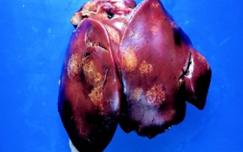
Circular, depressed saucer-shaped craters with raised yellowish borders in turkey liver parenchyma.
Swollen dark liver studded with diffuse pinpoint white necrotic micro-abscesses ('starry sky' appearance).
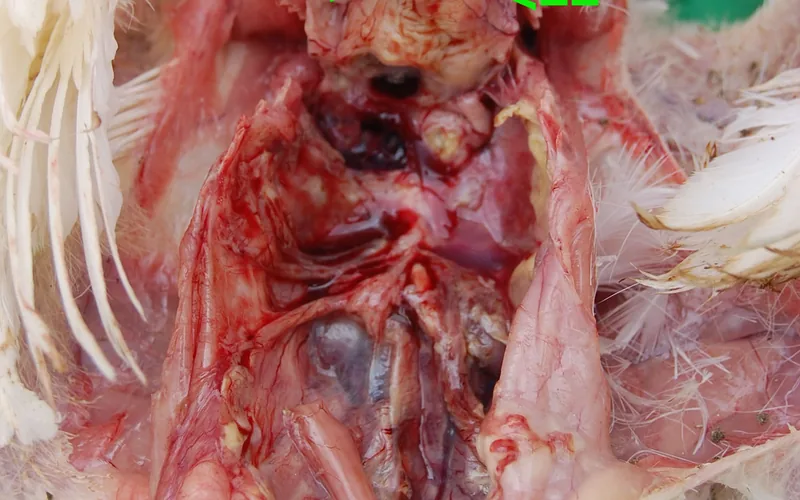
Thick yellow-white fibrinous pseudomembrane completely encasing the heart sac (frosted heart) and perihepatitis.

Massively enlarged, pale yellow-ochre friable liver with severe vascular rupture and fatal coelomic hematoma.
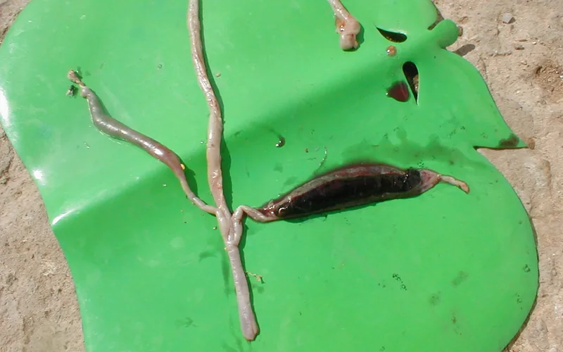
Opened blind ceca distended with frank coagulated blood, sloughed mucosa, and laminated fibrinous core.

Gas-distended friable small intestine lined with a diffuse, confluent tan-yellow 'Turkish towel' pseudomembrane.
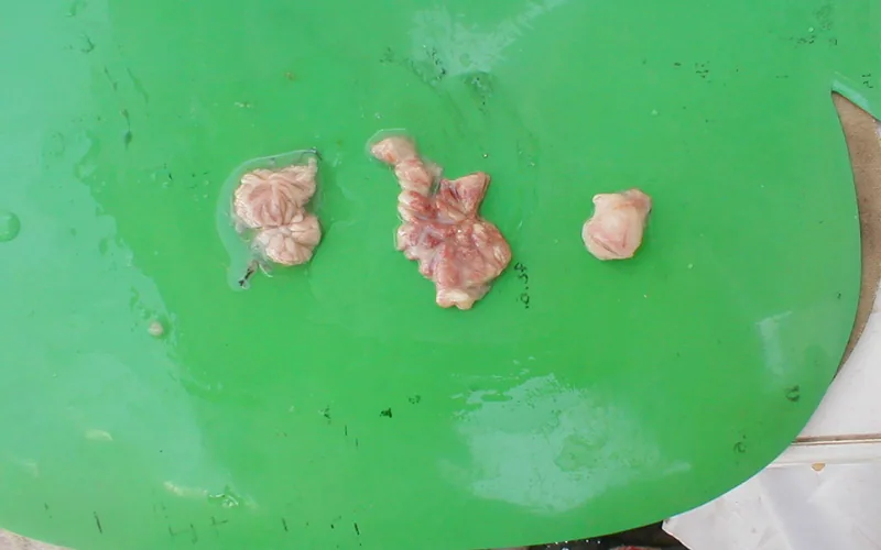
Direct post-mortem comparison: chronic atrophic bursa (left), acute edematous swelling (middle), and normal bursa (right).

Longitudinal section of chicken femur displaying severe pale fatty aplasia of hematopoietic bone marrow.

Diffuse subcutaneous petechiae and ecchymotic hemorrhages on unfeathered tarsometatarsal scales and footpads.
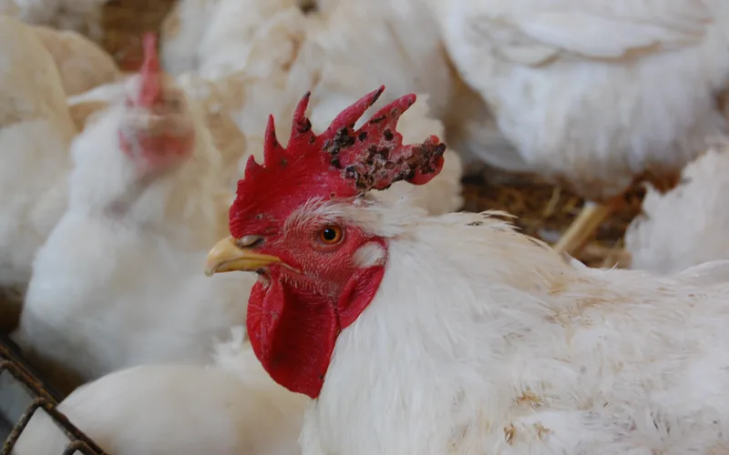
Proliferative nodular warty scabs firmly adhered to comb and facial skin; bleeding raw base upon removal.
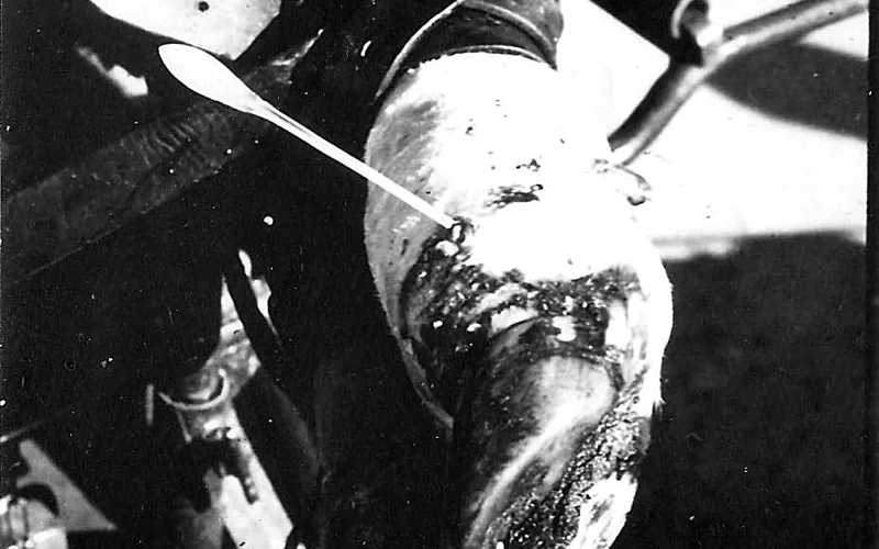
Creamy honey-like caseous exudate distending synovial sheaths of interdigital joints and hock tendon sheaths.
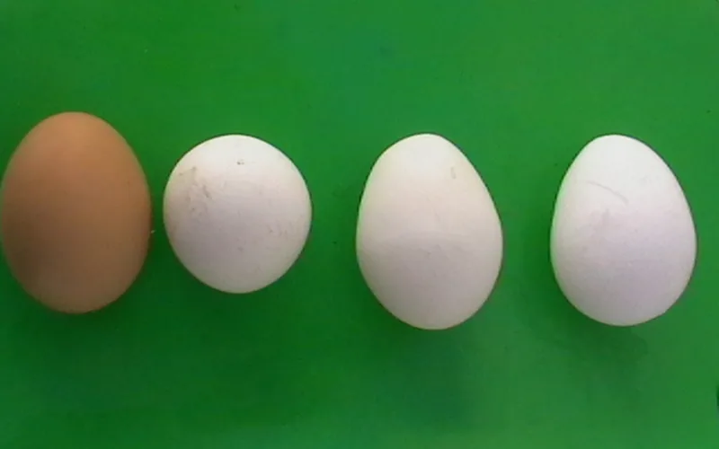
Thin-shelled, corrugated, ridged, and chalky eggs resulting from coronavirus viral hypoplasia of oviduct glands.
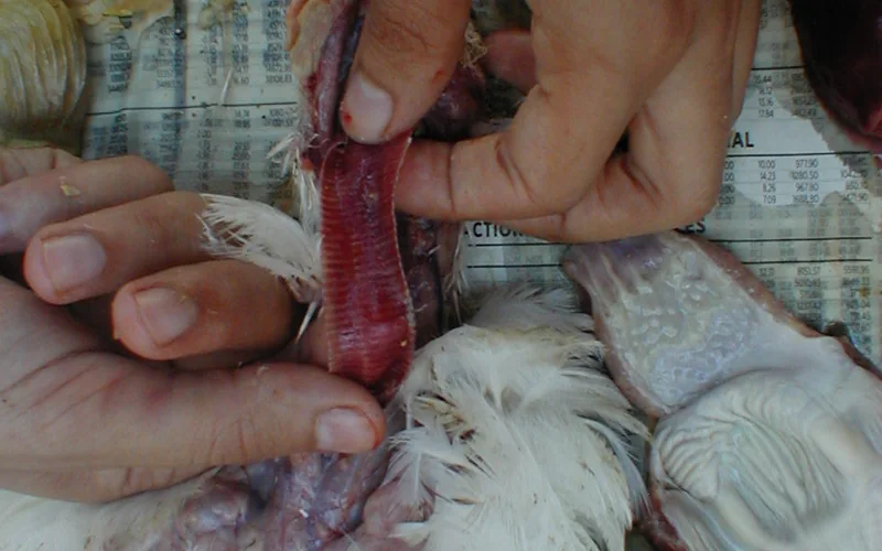
Opened broiler trachea displaying severe catarrhal-hemorrhagic mucosal inflammation, edema, and inflammatory exudate.
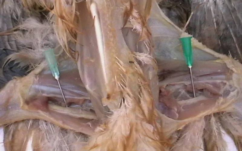
Comparative dissection displaying unilateral gross hypertrophy of the left sciatic nerve (2-3x enlargement) compared to normal right nerve.
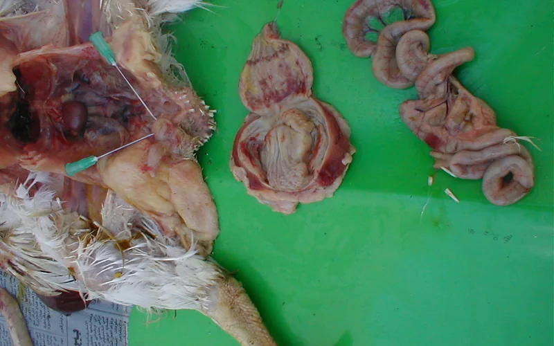
Longitudinally opened proventriculus displaying petechial to ecchymotic hemorrhages concentrated on mucosal papillae tips.
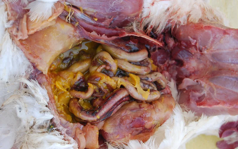
Coelomic cavity filled with inspissated cheesy yolk exudate, fibrinous peritonitis, and oviduct inflammation in mature hen.
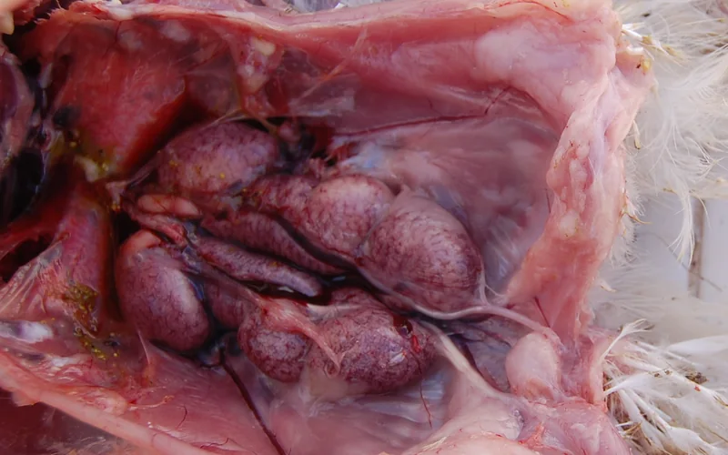
Enlarged pale lobulated kidneys distended with chalky white calcium urate crystal deposition in renal tubules.
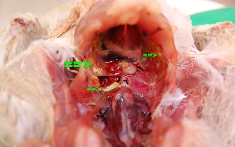
Thick, opaque yellow fibrinous exudate lining air sac membranes with loss of normal transparency.

Dark plum-colored congested lung parenchyma with pulmonary edema and bilateral cardiomegaly.

Opened visceral coelom displaying intense duodenal congestion, petechiae, and acute catarrhal enteritis.

Nodular cauliflower-like lymphoid/adenocarcinoma tumor masses expanding from the ovary through the abdominal cavity.

Severe inflammatory fibrinous exudate occluding the tracheal lumen and bifurcation causing acute asphyxiation.

Severe hepatic degeneration with pale yellowish necrotic foci and liver swelling in acute broiler autopsy.
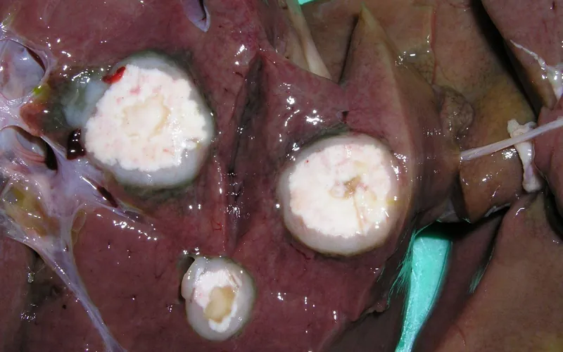
Multiple firm pale nodular granulomatous lesions expanding parenchymal tissue (Hjarre's disease).

Severe unilateral/bilateral infraorbital sinus distension, periorbital edema, chemosis, and closed eye.
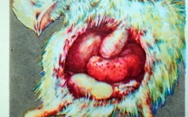
Enlarged mottled spleen (splenomegaly) with pinpoint focal white necrotic foci and subcapsular petechiae.
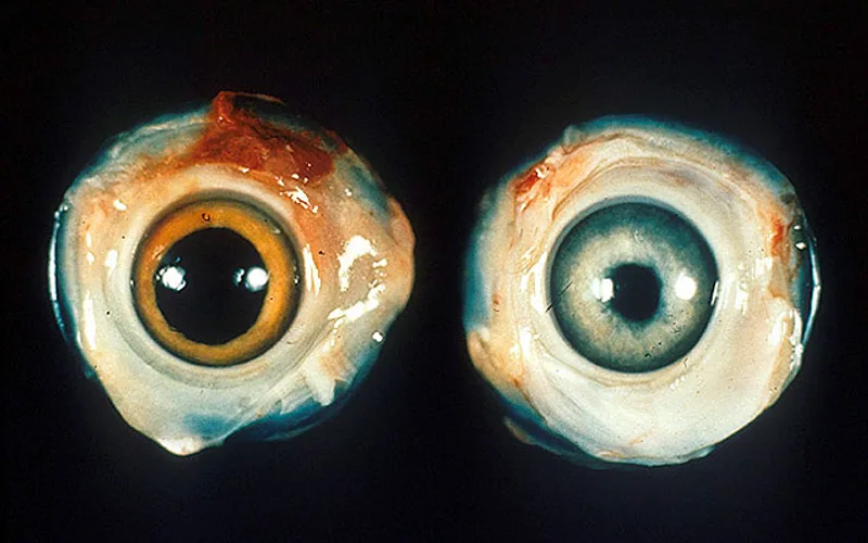
Mononuclear neoplastic infiltration into the iris causing loss of pigmentation ('grey eye') and irregular pupil.
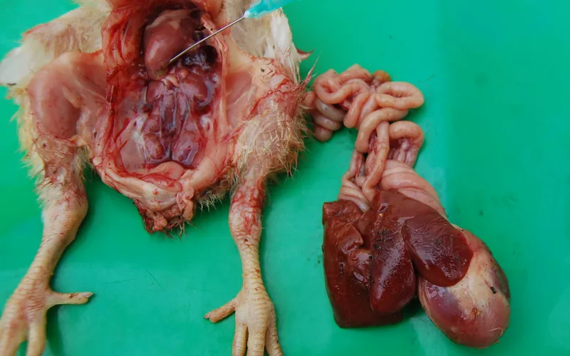
Distended translucent pericardial sac packed with amber straw-colored transudate ('hydropericardium syndrome').
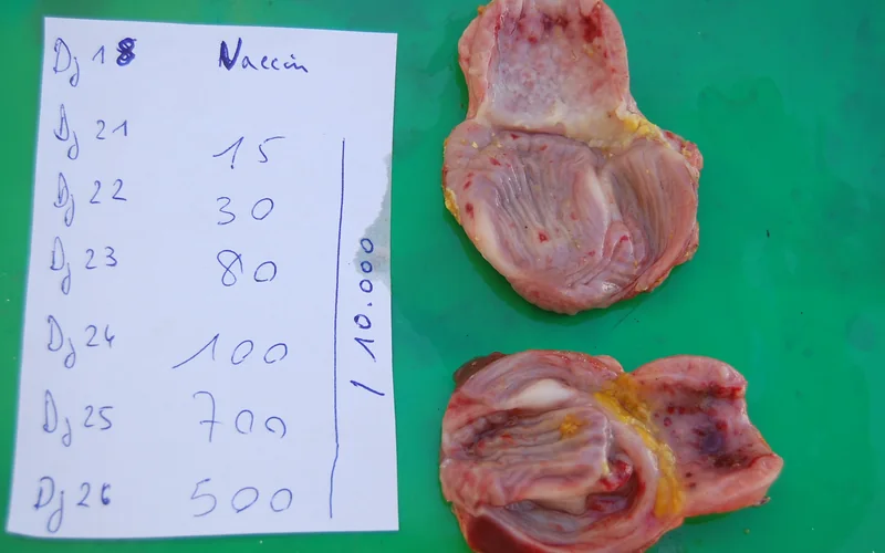
Prominent mucosal petechial hemorrhages beneath the koilin lining and proventricular-gizzard junction.
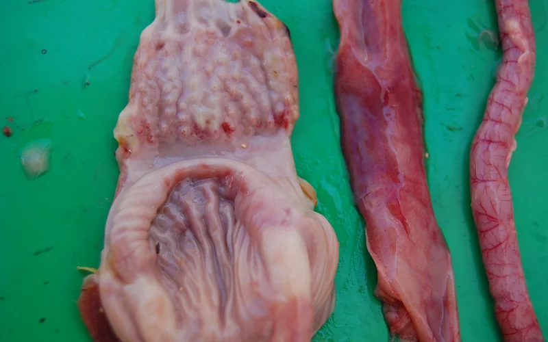
Raised button-like circular necrotic ulcers with hemorrhagic halos along the mucosal lining.
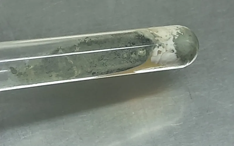
Multiple firm yellow-white to greenish disc-shaped mycotic granulomas (1-5 mm) throughout lung tissue.
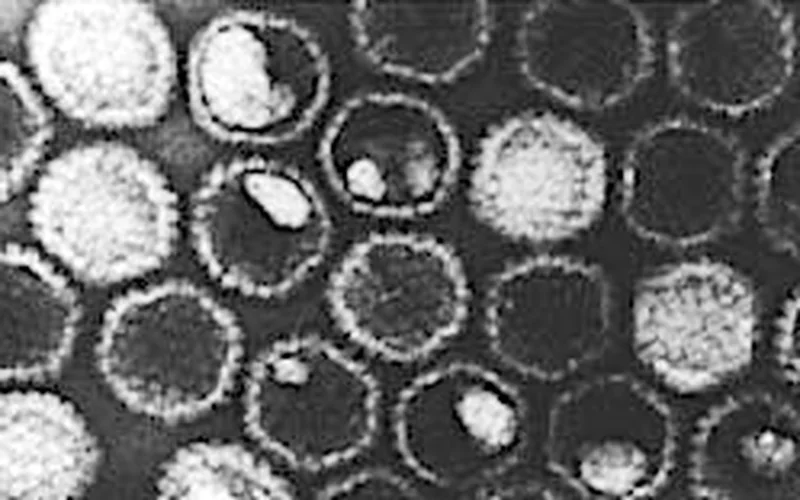
Transmission electron micrograph showing enveloped icosahedral Gallid alphaherpesvirus 1 virions.

Scanning electron micrograph of flask-shaped Mycoplasma gallisepticum with terminal bleb attachment organelle.
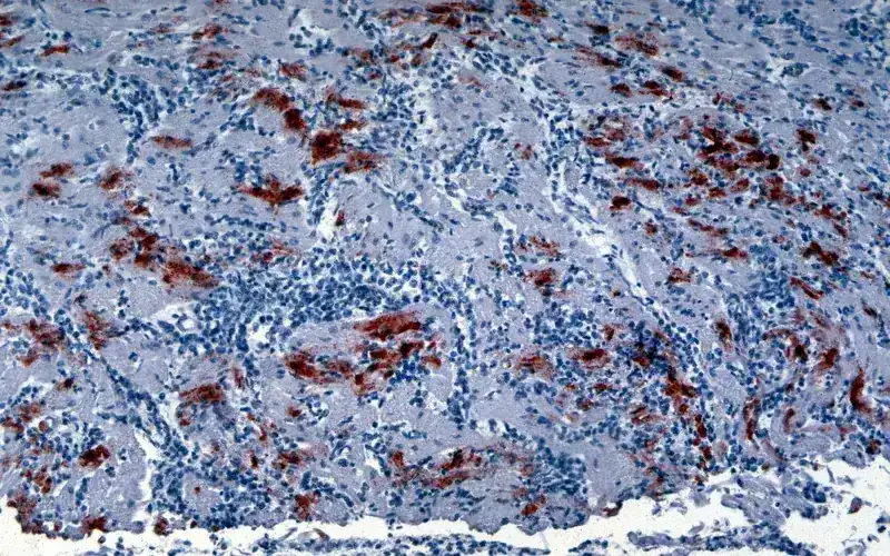
Severe prostration, bilateral wing drop, torticollis (twisted neck), and high mortality in infected flock.
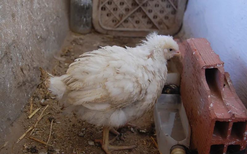
Huddled depressed broilers with ruffled plumage, pale combs, and blood-tinged watery droppings.
biotech Standard Operating Procedure for Systematic Avian Necropsy
To prevent diagnostic artifacts and avoid missing focal lesions, post-mortem examinations must proceed through four disciplined anatomical phases:
Examine eyes, nares, comb, wattles, and plumage. Check unfeathered shanks for subcutaneous petechiae (HPAI/vND). Disarticulate coxofemoral joints to lay the carcass stable on the necropsy tray.
Incise skin from vent to mandibular symphysis. Retract breast skin. Transect ribs at costochondral junctions with bone shears. Remove breastplate upward to inspect intact thoracic and abdominal air sacs.
Inspect liver capsule, pericardium, and heart. Transect proventriculus near thoracic inlet; reflect gastrointestinal tract posteriorly. Longitudinally open proventriculus, gizzard, and cecal tonsils.
Examine the Bursa of Fabricius dorsal to the cloaca. Dislodge kidneys from renal fossae to inspect sciatic plexus and roots for asymmetric hypertrophy and loss of transverse striations (Marek's Disease).
help_outline Avian Necropsy & Differential Pathology Clinical FAQs
How do you differentiate between Newcastle Disease and Highly Pathogenic Avian Influenza on post-mortem?
What is the hallmark gross difference between Marek's Disease and Avian Leukosis?
Why is the Johnson & Reid scoring system critical during gut coccidiosis necropsy?
What are the statutory reportable avian diseases requiring immediate veterinary authority notification?
Integrate necropsy pathology observations with our suite of precision veterinary calculators: